Review
. 2025 Feb 3;10(1):31.
doi: 10.1038/s41392-024-02071-0.

Homeostasis and metabolism of iron and other metal ions in neurodegenerative diseases

Affiliations
Review

Homeostasis and metabolism of iron and other metal ions in neurodegenerative diseases

Leilei Chen et al. Signal Transduct Target Ther. .

Abstract

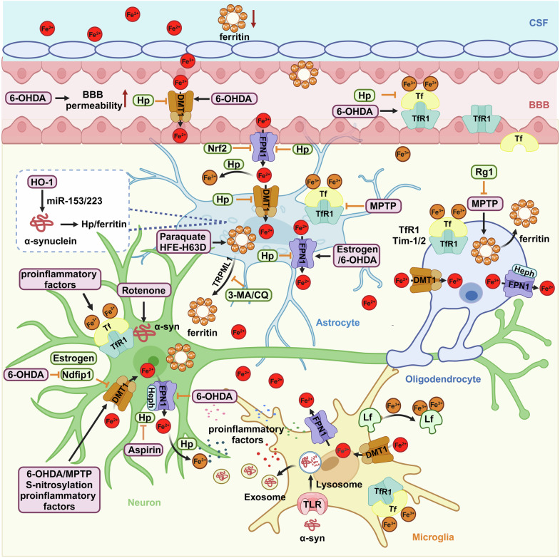
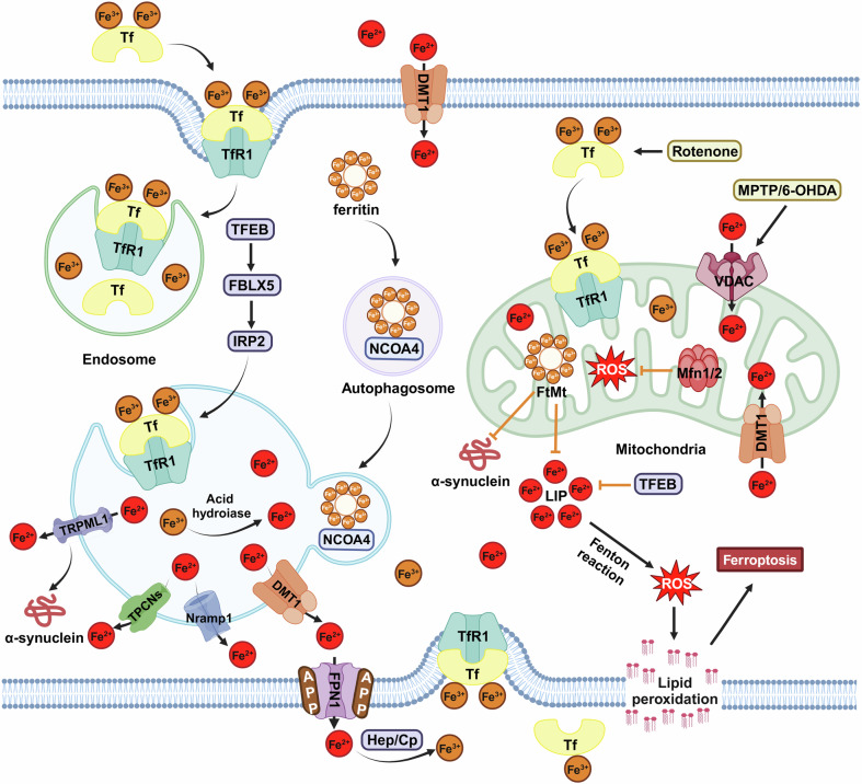
As essential micronutrients, metal ions such as iron, manganese, copper, and zinc, are required for a wide range of physiological processes in the brain. However, an imbalance in metal ions, whether excessive or insufficient, is detrimental and can contribute to neuronal death through oxidative stress, ferroptosis, cuproptosis, cell senescence, or neuroinflammation. These processes have been found to be involved in the pathological mechanisms of neurodegenerative diseases. In this review, the research history and milestone events of studying metal ions, including iron, manganese, copper, and zinc in neurodegenerative diseases such as Parkinson's disease (PD), Alzheimer's disease (AD), amyotrophic lateral sclerosis (ALS), and Huntington's disease (HD), will be introduced. Then, the upstream regulators, downstream effector, and crosstalk of mental ions under both physiologic and pathologic conditions will be summarized. Finally, the therapeutic effects of metal ion chelators, such as clioquinol, quercetin, curcumin, coumarin, and their derivatives for the treatment of neurodegenerative diseases will be discussed. Additionally, the promising results and limitations observed in clinical trials of these metal ion chelators will also be addressed. This review will not only provide a comprehensive understanding of the role of metal ions in disease development but also offer perspectives on their modulation for the prevention or treatment of neurodegenerative diseases.


Conflict of interest statement

Competing interests: The authors declare no competing interests.

Figures

Fig. 1
Fig. 1
Timeline and milestone events of study on iron and other metal ions in neurodegenerative diseases. The timeline begins at 1924 and expands to 2024. Milestone events of studying metal ions, including iron, manganese, copper, and zinc in neurodegenerative diseases, such as PD, AD, ALS, and HD, are listed in the figure. This figure was created with BioRender.com/d84k316
Fig. 2
Fig. 2
Downstream effectors of iron and other metal ions. a The import of iron mainly depends on TfR1-mediated endocytosis or DMT1, while FPN1 is the sole known exporter of iron. The import of copper mainly depends on CTR1, which can be upregulated by Cu(I)-GSH. DMT1 also participates in the import of copper. The export of copper is mediated by ATP7A/7B with the assistance of Atox1. As a copper-binding protein, Cp also functions as a ferroxidase to convert toxic ferrous iron into nontoxic ferric iron. Excessive copper leads to the aggregation of lipoylated proteins and the loss of iron-sulfur cluster proteins, resulting in cuproptosis. The import of manganese relies on the DAT, ZIP8, Tf/TfR system, or DMT1, while the export of manganese depends on SLC30A70 and FPN1. Manganese can activate ATM/ p53, which regulates cell cycles and reduces DNA damage. In the mitochondria, Mn2+ can bind to intermediate products of the TCA cycle and promote the generation of ROS, while Mn3+ can help SOD2 to mitigate the generation of ROS and prevent cells from undergoing apoptosis. ZIPs are the main channels for transporting zinc into the cytoplasm from extracellular or ER, while ZnTs are responsible for transporting zinc out of the cytoplasm or into synaptic vesicles, lysosomes, and Golgi. Both ferrous iron and copper can promote the Fenton reaction, leading to the generation of ROS and initiating oxidative stress. However, zinc can compete with copper or iron, thereby preventing the generation of ROS. Both excessive Fe2+ and Mn2+ iron can lead to the accumulation of lipid peroxidation and trigger ferroptosis, which can be inhibited by copper chelators. b High levels of metal ions, including iron, manganese, copper, and zinc have been found to be involved in cell senescence. This process can be rescued by the iron chelator DFO. c Excessive metal ions such as iron, manganese, copper, and zinc can also activate microglia and astrocytes to release pro-inflammatory cytokines, thereby triggering neuroinflammation. This figure was created with BioRender.com/j25f189
Fig. 3
Fig. 3
Iron dysregulation among different cells occurs in the state of PD. The increased iron level and decreased ferritin level have been found in the CSF under the state of PD. The increased permeability of BBB facilitates the import of iron through TfR1 by microvascular endothelial cells, which is then released into the brain through FPN1. Once iron crosses the BBB, astrocytes absorb it and mediate its transfer to neurons. Astrocytes have shown a neuroprotective role in PD through the secretion of hepcidin and ferritin, which not only decreases brain iron load by regulating FPN1 on microvascular endothelial cells but also controls iron import by regulating iron-related proteins. With exposure to α-synuclein, iron, or neurotoxins, astrocytes also exhibit an iron-releasing phenotype that is detrimental to neighboring neurons. Microglia are the most susceptible neuronal cells to ferroptosis. Upon stimulation, activated microglia release proinflammatory factors, iron, as well as α-synuclein, which interact and exacerbate dopaminergic neurotoxicity. On the other hand, activated microglia can synthesize lactoferrin and protect vulnerable dopaminergic neurons. Oligodendrocytes harbor the highest concentration of iron in the CNS, which can secrete a ferritin heavy chain and protect dopaminergic neurons from oxidative stress. Abnormal import and export of iron-mediated by iron-related proteins cause iron deposition in nigral dopaminergic neurons, resulting in the loss of dopaminergic neurons in PD. This figure was created with BioRender.com/f23j195
Fig. 4
Fig. 4
Iron dysregulation in the lysosome and mitochondria of PD. The Tf-TfR system mediates the uptake of Fe3+ and DMT1 mediates the uptake of Fe2+, which are the two major pathways for the neuronal iron influx. FPN1 is responsible for exporting Fe2+ with the help of APP, Cp, or hephaestin (Hep). Endosome contains abundant Fe3+ through Tf-TfR1 uptake, while autophagosome contains ferritin bound to NCOA4, which are the main source of iron in lysosomes. In the acidic and reducing environment of the lysosome, Fe3+ is reduced to Fe2+ and released into the cytosolic through DMT1, TRPML1, Nramp1, or TPCNs. The overexpression of TFEB can upregulate the synthesis of TfR1 through the FBXL5-IRP2 pathway and increase the localization of TfR1 in lysosomes, thereby facilitating the import and temporary storage of iron in lysosomes. VDAC, Tf-TfR2, and DMT1, all of which are located on the outer mitochondrial membrane, are responsible for the iron transport across the outer mitochondrial membrane. The transport of iron across the inner mitochondrial membrane is mediated by Mfrn1/2. In the mitochondrial matrix, iron is stored in FtMt. Rotenone induces an increased level of Tf, while MPTP or 6-OHDA induces increased levels of VDAC in mitochondria. Additionally, excessive iron induces the generation of ROS and lipid peroxidation through the Fenton reaction, further leading to ferroptosis. However, overexpression of TFEB or FtMt has the ability to suppress α-synuclein aggregation and decrease the cellular labile iron pool. All these processes contribute to the nigral dopaminergic neuronal death. This figure was created with BioRender.com/u48z218
Fig. 5
Fig. 5
Crosstalk between dysregulation of metal ions and pathological proteins in neurodegenerative diseases. Excessive iron affects α-synuclein aggregation, while misfolded α-synuclein accelerates iron deposition. There are two copper-binding sites in the N-terminus of α-synuclein that increase its aggregation. Silencing CTR1 can decrease the phosphorylation and aggregation of α-synuclein. PARK9 deficiency induces increased zinc levels and α-synuclein aggregation. Manganese can indirectly stimulate the expression of α-synuclein through activating the ERK1/2 MAPK pathway. On the other hand, α-synuclein aggregation could enhance manganese-induced neurotoxicity through the NF-κB pathway. The binding of IRP to iron inhibits the IRE, which increases the expression of APP. APP can be cleaved by β- and γ-secretase in early endosomes to form Aβ in the amyloidogenic pathway. On one hand, excessive metal ions induce increased levels of Aβ monomers; on the other hand, metal ions can bind to Aβ and promote its aggregation. Iron, copper, and zinc enhance tau phosphorylation by activating CDK5, GSK-3β, or MAPKs and inactivating PP-2A activity to accelerate the formation of NFTs. Manganese exposure promotes the development of ALS. Additionally, partial deficiency of SOD2 significantly exacerbates motor deficits. Iron dysregulation induced by SOD1G93A is mediated by impairment of the Akt/FOXO3a signaling pathway. Meanwhile, increased levels of copper, zinc, and metallothioneins-I/II/III are observed in SOD1G93A mice. Copper has the ability to bind with the N-terminus of HTT proteins, thereby promoting their aggregation. Mutant HTT can lead to a deficiency in neuronal manganese, which affects arginase activity. Additionally, mutant HTT inhibits the binding of Sp1 to the promoter of the ZnT3 gene, resulting in decreased levels of synaptic vesicular zinc in the hippocampus, cortex, and striatum. This figure was created with BioRender.com/q53d457

References

    1. Lhermitte, J., Kraus, W. M. & McAlpine, D. Original Papers: On the occurrence of abnormal deposits of iron in the brain in parkinsonism with special reference to its localisation. J. Neurol. Psychopathol.5, 195–208 (1924). - PMC - PubMed
    1. Dexter, D. T. et al. Increased nigral iron content in postmortem parkinsonian brain. Lancet2, 1219–1220 (1987). - PubMed
    1. Dexter, D. T. et al. Alterations in the levels of iron, ferritin and other trace metals in Parkinson’s disease and other neurodegenerative diseases affecting the basal ganglia. Brain114, 1953–1975 (1991). - PubMed
    1. Dexter, D. T. et al. Increased nigral iron content and alterations in other metal ions occurring in brain in Parkinson’s disease. J. Neurochem52, 1830–1836 (1989). - PubMed
    1. Mann, V. M. et al. Complex I, iron, and ferritin in Parkinson’s disease substantia nigra. Ann. Neurol.36, 876–881 (1994). - PubMed

MeSH terms