. 2025 Jan;17(1):77-93.
doi: 10.4168/aair.2025.17.1.77.

Inotodiol Attenuates Mucosal Inflammation in a Mouse Model of Eosinophilic Chronic Rhinosinusitis

Affiliations

Inotodiol Attenuates Mucosal Inflammation in a Mouse Model of Eosinophilic Chronic Rhinosinusitis

Jaein Chung et al. Allergy Asthma Immunol Res. 2025 Jan.

Abstract

Purpose: Inotodiol (22-hydroxy lanosterol), a unique component of chaga mushrooms, is believed to be a medicinal component with reported antitumor, antiviral, and anti-inflammatory properties. This study evaluated the therapeutic potential and underlying mechanisms of inotodiol in eosinophilic chronic rhinosinusitis (ECRS).

Methods: An ECRS mouse model was established using female BALB/c mice. Forty mice were categorized into 4 groups: the control group (n = 10), ECRS group treated with solvent (n = 10), ECRS group treated with inotodiol 20 mg/kg (n = 10), and ECRS group treated with dexamethasone 10 mg/kg (n = 10). The nasal lavage fluid and tissue samples from mice were analyzed for cytokine and chemokine expression as well as for the severity of mucosal inflammation. Enzyme-linked immunosorbent assay, quantitative reverse transcription-polymerase chain reaction, histopathological staining, and immunofluorescence techniques were employed. The human eosinophil cell line (EoL-1) and dispersed nasal polyp cells (DNPCs) were used to assess inotodiol-induced eosinophil apoptosis in vitro via immunofluorescence, flow cytometry, and proteome profiler antibody array analysis.

Results: Inotodiol significantly reduced the secretion of T2 cytokine and mast cell tryptase as well as the expression of Th cytokines, chemokines, and proinflammatory/inflammatory cytokines in ECRS mice. Furthermore, it suppressed mucosal inflammatory features such as polyp formation, epithelial thickening, and eosinophil infiltration. Inotodiol treatment reduced mast cell activation and increased eosinophil apoptosis in the nasal mucosa of ECRS mice. Notably, inotodiol also induced apoptosis in EoL-1 cells and DNPCs, which may contribute to its anti-inflammatory effects.

Conclusions: Inotodiol could be a potential therapeutic agent for ECRS by modulating immune responses and reducing mucosal inflammation.

Keywords: Apoptosis; cytokines; eosinophils; inflammation; inotodiol; mushroom; nasal polyp; rhinosinusitis.


Conflict of interest statement

There are no financial or other issues that might lead to conflict of interest.

Figures

Fig. 1
Fig. 1. Experimental protocol for developing a murine model of eosinophilic chronic rhinosinusitis with nasal polyposis. Mice were intraperitoneally sensitized on days 0 and 5 with either 2 mg of aluminum hydroxide alone or 2 mg of aluminum hydroxide combined with 25 mg of OVA, followed by intranasal challenge with 3% OVA 5 times in week 3. The mice received 3% OVA and SEB from weeks 4 to 8, and from weeks 8 to 12, respectively, intranasally 3 times a week. Forty mice were divided into 4 groups: the CON group (n = 10), NP group (Tween 80% + 0.9% NaCl, n = 10), NP + Ino (n = 10), and NP + Dex group (n = 10). The CON group received phosphate-buffered saline instead of OVA, SEB, inotodiol, or dexamethasone. The NP, NP + Ino, and NP + Dex groups received intraperitoneal injections of 20 µL of solvent, 20 mg/kg inotodiol, and 10 mg/kg dexamethasone, respectively, from weeks 8 to 12.
OVA, ovalbumin; SEB, staphylococcal enterotoxin B; I.N., intranasal; I.P, intraperitoneal; CON, control group; NP, nasal polyp with solvent treatment group; NP + Ino, nasal polyp with inotodiol treatment group; NP + Dex, nasal polyp with dexamethasone treatment group.
Fig. 2
Fig. 2. Comparison of IL-4, IL-5, and MCT production in the nasal lavage fluid between the experimental groups. (A–C) Levels of IL-4 (A), IL-5 (B), and MCT (C) were measured using enzyme-linked immunosorbent assay (n = 8). The NP group showed significantly higher levels of IL-4, IL-5, and MCT than in the control, NP + Ino, and NP + Dex groups.
IL, interleukin; CON, control group; NP, nasal polyp with solvent treatment group; NP + Ino, nasal polyp with inotodiol treatment group; NP + Dex, nasal polyp with dexamethasone treatment group; MCT, mast cell tryptase. *P < 0.01, P < 0.001.
Fig. 3
Fig. 3. Comparison of the mRNA expression of cytokines and chemokines in the nasal mucosa between experimental groups. (A–F) The expression levels of IL-4, IL-5, IL-10, IL-13, IL-17A, and IFN-γ were significantly higher in the NP group than in the CON, NP + Ino, and NP + Dex groups. (G–J) Similarly, the expression of IL-6, IL-1β, IL-25, and IL-33 was significantly elevated in the NP group compared to the CON, NP + Ino, and NP + Dex groups. (K–M) The expression of chemokines, including CCL1, CCL2, and CXCL2, was also significantly higher in the NP group than in the CON, NP + Ino, and NP + Dex groups.
IL, interleukin; CON, control group; NP, nasal polyp with solvent treatment group; NP + Ino, nasal polyp with inotodiol treatment group; NP + Dex, nasal polyp with dexamethasone treatment group; IFN, interferon; CCL, C-C motif chemokine ligand; CXCL2, chemokine (C-X-C motif) ligand 2. *P < 0.05, P < 0.01, P < 0.001, §P < 0.0001.
Fig. 4
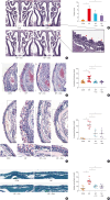
Fig. 4. Histopathologic analysis of the sinonasal mucosa across experimental groups. (A) Representative images of H&E staining, with polyp counts indicated by asterisks, in the sinonasal cavity for each group. (B) The number of polyps in the nasal cavity was significantly lower in the NP + Dex and CON groups than in the NP group. (C) Epithelial thickness was evaluated using H&E staining. (D) Epithelial thickness in the sinonasal mucosa was significantly reduced in the NP + Ino and NP + Dex groups compared to the NP group. (E) Eosinophil infiltration (arrowheads) in the sinonasal mucosa was assessed using Congo red staining. (F) The number of infiltrated eosinophils in the nasal mucosa was significantly lower in the NP + Ino and NP + Dex groups than in the NP group. (G) Goblet cells in the sinonasal epithelium were examined using periodic acid-Schiff staining. (H) The number of goblet cells was significantly reduced in the NP + Ino and NP + Dex groups compared to the NP group. (I) The morphology and degranulation of mast cells were evaluated using toluidine blue staining, revealing both degranulation and activation in the NP group. (J) Representative immunofluorescence images of mast cell tryptase in each group. (K) Tryptase expression was significantly higher in the NP group than in the NP + Ino and NP + Dex groups. Scale bar: 50 μm.
CON, control group; NP, nasal polyp with solvent treatment group; NP + Ino, nasal polyp with inotodiol treatment group; NP + Dex, nasal polyp with dexamethasone treatment group; H&E, hematoxylin and eosin. *P < 0.05, P < 0.01, P < 0.0001.
Fig. 5
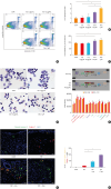
Fig. 5. Effects of inotodiol treatment on eosinophil apoptosis. (A) Apoptosis in PBMCs treated with inotodiol was measured via flow cytometric analysis using FITC annexin V staining. (B) The number of apoptotic cells was significantly higher only in the PBMCs treated with Dex. (C) Toxicity of the reagents was assessed based on LDH release in the supernatant; no significant differences in LDH release percentages were observed between the groups. (D) EoL-1 cells were stimulated with BA (1 mM) for 24 h, followed by inotodiol treatment (5 and 10 μg/mL) or Dex (1 μM) for an additional 24 h. Morphological changes in eosinophils were observed using H&E staining, revealing increased cytoplasmic blebbing and nuclear fragmentation in inotodiol- and Dex-treated eosinophils (arrowheads). (E) Proteome profiler analysis demonstrated that the expression of pro-apoptotic and apoptotic signaling pathway molecules, including cleaved caspase 3, was significantly increased in EoL-1 cells treated with inotodiol (10 µg/mL) compared to those treated with media alone. (F) Expression of cleaved caspase 3 in eosinophils was examined in the nasal mucosa of mice with eosinophilic chronic rhinosinusitis using a double immunofluorescence assay. (G) The number of double-positive cells (cleaved caspase 3 and Siglec-F) was significantly higher in the NP + Ino and NP + Dex groups than in the NP group. Scale bar: 25 μm.
CON, control group; Dex, dexamethasone; BA, butyric acid; NP, nasal polyp with solvent treatment group; NP + Ino, nasal polyp with inotodiol treatment group; NP + Dex, nasal polyp with dexamethasone treatment group; DAPI, 4′,6-diamidino-2-phenylindole; PBMC, peripheral blood mononuclear cell; LDH, lactate dehydrogenase. *P < 0.05, P < 0.01, P < 0.001, §P < 0.0001.
Fig. 6
Fig. 6. Inotodiol treatment induced eosinophil apoptosis and suppressed IL-13 production in DNPCs from ECRS patients. (A) Representative immunofluorescence staining of DNPC cultures from ECRS patients showing DAPI (blue), cleaved caspase 3 (green), MBP (red), and signal overlay (yellow). (B) The graph depicts the percentage of apoptotic eosinophils (cleaved caspase 3 + MBP) in the MBP-positive stained area of each culture, indicating a significant increase in apoptotic eosinophils in inotodiol-treated DNPCs compared to those treated with media alone from ECRS patients (n = 5). (C) The production of IL-13 in the DNPC lysate was significantly suppressed in INO-treated DNPCs harvested from ECRS patients compared to those from non-ECRS patients. Scale bar: 50 μm. The P value was calculated using Fisher’s least significant difference method.
MBP, major basic protein; DAPI, 4′,6-diamidino-2-phenylindole; CON, solvent treatment; SEB, staphylococcus enterotoxin B; INO, inotodiol; IL, interleukin; ECRS, eosinophilic chronic rhinosinusitis; NECRS, non-eosinophilic chronic rhinosinusitis; DNPC, dispersed nasal polyp cell. *P < 0.05, P < 0.01.

References

    1. Ahn JC, Kim JW, Lee CH, Rhee CS. Prevalence and risk factors of chronic rhinosinusitus, allergic rhinitis, and nasal septal deviation: results of the Korean National Health and Nutrition Survey 2008–2012. JAMA Otolaryngol Head Neck Surg. 2016;142:162–167. - PubMed
    1. Hastan D, Fokkens WJ, Bachert C, Newson RB, Bislimovska J, Bockelbrink A, et al. Chronic rhinosinusitis in Europe--an underestimated disease. A GA²LEN study. Allergy. 2011;66:1216–1223. - PubMed
    1. Bachert C, Marple B, Schlosser RJ, Hopkins C, Schleimer RP, Lambrecht BN, et al. Adult chronic rhinosinusitis. Nat Rev Dis Primers. 2020;6:86. - PubMed
    1. Wang X, Zhang N, Bo M, Holtappels G, Zheng M, Lou H, et al. Diversity of TH cytokine profiles in patients with chronic rhinosinusitis: a multicenter study in Europe, Asia, and Oceania. J Allergy Clin Immunol. 2016;138:1344–1353. - PubMed
    1. Stevens WW, Peters AT, Tan BK, Klingler AI, Poposki JA, Hulse KE, et al. Associations between inflammatory endotypes and clinical presentations in chronic rhinosinusitis. J Allergy Clin Immunol Pract. 2019;7:2812–2820.e3. - PMC - PubMed