. 2025 Jan 14;10(3):2628-2639.
doi: 10.1021/acsomega.4c07666. eCollection 2025 Jan 28.

Intracellular Biomacromolecule Delivery by Stimuli-Responsive Protein Vesicles Loaded by Hydrophobic Ion Pairing

Affiliations

Intracellular Biomacromolecule Delivery by Stimuli-Responsive Protein Vesicles Loaded by Hydrophobic Ion Pairing

Mikaela A Gray et al. ACS Omega. .

Abstract

Proteins can perform ideal therapeutic functions. However, their large size and significant surface hydrophilicity and charge prohibit them from reaching intracellular targets. These chemical features also render them poorly encapsulated by nanoparticles used for intracellular delivery. In this work, a novel combination of protein vesicles and hydrophobic ion pairing (HIP) was used to load protein cargo and achieve cytosolic delivery to overcome the limitations of previous protein vesicle properties. Protein vesicles are thermally self-assembling nanoparticles made from elastin-like polypeptide (ELP) fused to an arginine-rich leucine zipper and a globular protein fused to a glutamate-rich leucine zipper. To impart stimuli-responsive disassembly, physiological stability, and small size, the ELP sequence was modified to include histidine and tyrosine residues. HIP was used to load and release protein cargo requiring endosomal escape for cytosolic function. HIP vesicles enabled delivery of cytochrome c, a cytosolically active protein, and a significant reduction in viability in both a traditional two-dimensional (2D) human cancer cell line culture and a biomimetic three-dimensional (3D) organoid model of acute myeloid leukemia. By examining the uptake of positively and negatively charged fluorescent protein cargos loaded by HIP, this work revealed the necessity of HIP for cytosolic cargo delivery and how HIP loading influences protein vesicle self-assembly and disassembly using microscopy, small-angle X-ray scattering, and nanoparticle tracking analysis. HIP protein vesicles have the potential to broaden the use of intracellular proteins as therapeutics for various diseases and extend protein vesicles to deliver other biomacromolecules, as the strategy developed here resulted in the first cytosolic protein cargo delivery using protein vesicles.


Conflict of interest statement

The authors declare no competing financial interest.

Figures

Figure 1
Figure 1
Protein vesicle characterization. (A) Illustration of self-assembling protein building blocks composed of mCherry-ZE and a mixture of ZR-ELPs using a 0.3 mCherry-ZE/ZR-ELP ratio and 30 μM total ELP using a 12 Y5-ZR-ELP (labeled with yellow spheres):1 H15-ZR-ELP (labeled with brown spheres) molar ratio with 0.15 M NaCl. (B) DLS analysis showing the size distribution of vesicles and stability of vesicles when diluted to 70% of original concentration into PBS. (C) Epifluorescence image of structures composed of mCherry-ZE and a mixture of ZR-ELPs using a 0.05 ZE/ZR ratio for enhanced visualization and 30 μM total ELP using a 12 Y5-ZR-ELP: 1 H15-ZR-ELP molar ratio in 0.15 M NaCl. The image was digitally enlarged 10×.
Figure 2
Figure 2
CytC vesicle characterization and delivery. (A) TEM of HIP CytC OA-loaded vesicles composed of mCherry-ZE and a mixture of ZR-ELPs using a 0.3 ZE/ZR ratio, 30 μM total ELP with a 12 Y5-ZR-ELP: 1 H15-ZR-ELP molar ratio, 10 μM CytC cargo, and 0.15 M NaCl. (B) DLS analysis showing the size distribution of cargo-loaded vesicles. (C) HeLa cell viability measured by an MTT assay 48 h after treatment with 2 μM CytC relative to media control. (D) Vesicle diffusion within an acute myeloid leukemia organoid. (E) Acute myeloid leukemia bone marrow organoid K562 cell viability 24 h after treatment with 2 μM CytC or 6 μM Dox showing the ability to enter the cytosol of cells in a tumor-like environment. One-way ANOVA was utilized with p > 0.05 n.s., p < 0.001***, p < 0.0001****, and n = 3 groups with each experiment repeated at least twice. Error bars are standard deviation from the mean.
Figure 3
Figure 3
Empty, sfGFP(−10), and sfGFP(+10) loaded vesicle characterization. (A) DLS size distribution of cargo-loaded vesicles composed of mCherry-ZE and a mixture of ZR-ELPs using a 0.3 ZE/ZR ratio, 30 μM total ELP using a 12 Y5-ZR-ELP: 1 H15-ZR-ELP molar ratio with and without 10 μM of sfGFP(−10) cargo in 0.15 M NaCl or with HIP sfGFP(−10) BA. (B) Epifluorescence images of structures composed of larger vesicles using a 0.05 ZE/ZR ratio (all other conditions are the same as (A) for cargo visualization). Scale bars are 0.5 μm, and images were digitally magnified 100×. (C) DLS size distribution of cargo-loaded vesicles composed of mCherry-ZE and a mixture of ZR-ELPs using a 0.3 ZE/ZR ratio and 30 μM total ELP using a 12 Y5-ZR-ELP: 1 H15-ZR-ELP molar ratio with and without 10 μM of sfGFP(+10) cargo in 0.15 M NaCl or with HIP sfGFP(+10) OA or SD. (D) Epifluorescence images of structures composed of larger vesicles using a 0.05 ZE/ZR ratio (all other conditions are the same as (A) for cargo visualization). Scale bars are 0.5 μm, and images were digitally magnified 100×.
Figure 4
Figure 4
Effect of cargo and HIP on vesicle transition temperatures, Tt. Transition temperatures of empty, sfGFP(−10), and sfGFP(−10) BA HIP vesicles with 10 μM sfGFP(−10) were determined from the midpoint of each linear region of the turbidity profile.
Figure 5
Figure 5
sfGFP(−10) and sfGFP(+10) loaded vesicle uptake and cellular viability. (A) Flow cytometry analysis showing median cell fluorescence of HeLa cells incubated with 1 μM sfGFP(−10) cargo for 24 h. (B) Viability 24 h after treatment using the MTT assay relative to media control. (C) Flow cytometry analysis showing median cell fluorescence of HeLa cells incubated with 1 μM sfGFP(+10) cargo for 24 h. (D) Viability 24 h after treatment using the MTT assay relative to media control. (E) Viability 48 h after treatment with 2 μM sfGFP(+10) loaded with 1 sfGFP(+10):16 OA and 1 sfGFP(+10):13 SD charge ratio using the MTT assay relative to media control. One-way ANOVA was utilized with p > 0.05 n.s., p < 0.05*, p < 0.01**, p < 0.0001****, and n = 3 groups with each experiment repeated at least twice. Error bars are standard deviation from the mean.
Figure 6
Figure 6
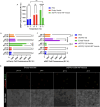
Energy-dependent endocytosis and RBC membrane interactions of sfGFP(−10) loaded vesicles. (A) Energy-dependent HeLa cell uptake using flow cytometry analysis of median fluorescence after 24 h of incubation with 1 μM sfGFP(−10). (B, C) RBC median fluorescence due to binding interactions with vesicles (B, mCherry fluorescence) or cargo (C, sfGFP fluorescence) at physiological and endosomal pH. 0.5% v/v RBCs were treated for 1 h, washed, and analyzed using flow cytometry and microscopy (scale bar: 20 μm). (D) Vesicles were composed of mCherry-ZE and a mixture of ZR-ELPs using a 0.3 ZE/ZR ratio and 30 μM total ELP with a 12 Y5-ZR-ELP: 1 H15-ZR-ELP molar ratio and 10 μM sfGFP(−10) cargo in 0.15 M NaCl and then treated according to the EE (using 0.413 or 0.51 mg/mL total vesicle proteins) to HeLa or RBC at 1 μM sfGFP. Two-way ANOVA was utilized with p > 0.05 n.s., p < 0.001***, p < 0.0001****, and n = 3 groups with each experiment repeated at least twice. Error bars are standard deviation from the mean.

Similar articles

Cited by

References

    1. Mocellin S.; Rossi C. R.; Pilati P.; Nitti D. Tumor Necrosis Factor, Cancer and Anticancer Therapy. Cytokine & Growth Factor Reviews 2005, 16 (1), 35–53. 10.1016/j.cytogfr.2004.11.001. - DOI - PubMed
    1. Roberts T. C.; Langer R.; Wood M. J. A. Advances in Oligonucleotide Drug Delivery. Nat. Rev. Drug Discovery 2020, 19 (10), 673–694. 10.1038/s41573-020-0075-7. - DOI - PMC - PubMed
    1. Porello I.; Cellesi F. Intracellular Delivery of Therapeutic Proteins. New Advancements and Future Directions. Front. Bioeng. Biotechnol. 2023, 11, 121179810.3389/fbioe.2023.1211798. - DOI - PMC - PubMed
    1. Leader B.; Baca Q. J.; Golan D. E. Protein Therapeutics: A Summary and Pharmacological Classification. Nat. Rev. Drug Discovery 2008, 7 (1), 21–39. 10.1038/nrd2399. - DOI - PubMed
    1. He C.; Tang Z.; Tian H.; Chen X. Co-Delivery of Chemotherapeutics and Proteins for Synergistic Therapy. Advanced drug delivery reviews 2016, 98, 64–76. 10.1016/j.addr.2015.10.021. - DOI - PubMed

LinkOut - more resources