. 2025 Jan 17:15:1459401.
doi: 10.3389/fmicb.2024.1459401. eCollection 2024.

Characterization of three novel Helicobacter species infecting stomachs of dogs and cats: Helicobacter gastrocanis sp. nov., Helicobacter gastrofelis sp. nov., and Helicobacter felistomachi sp. nov

Affiliations

Characterization of three novel Helicobacter species infecting stomachs of dogs and cats: Helicobacter gastrocanis sp. nov., Helicobacter gastrofelis sp. nov., and Helicobacter felistomachi sp. nov

Emiko Rimbara et al. Front Microbiol. .

Abstract

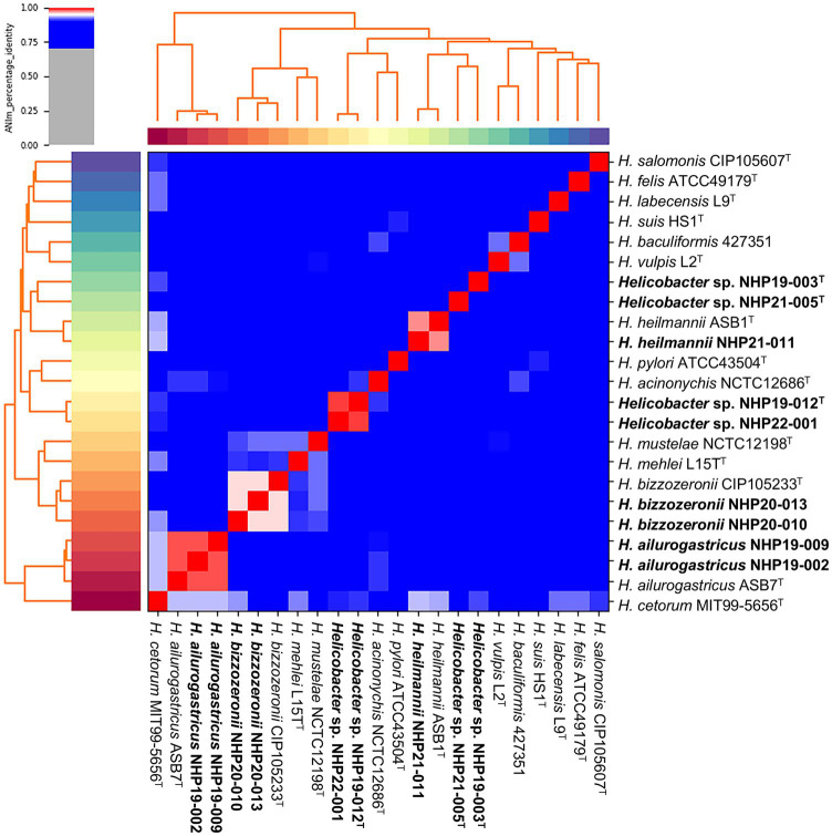
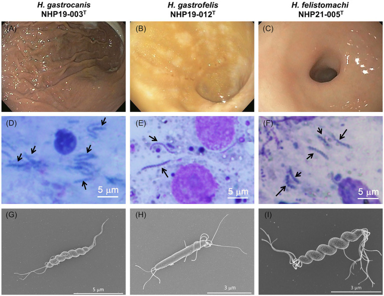
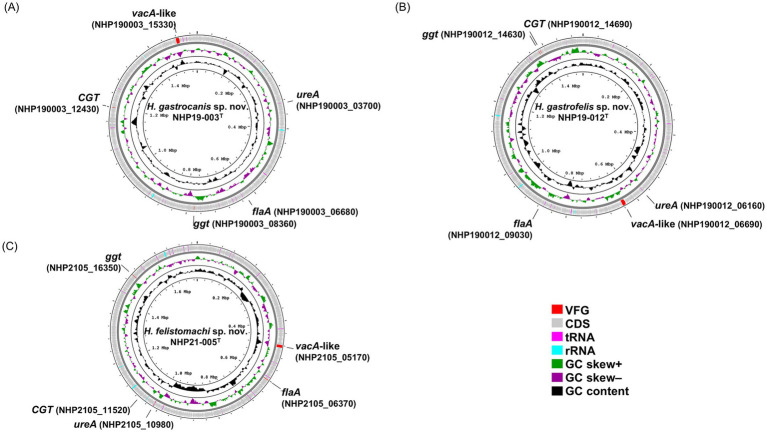
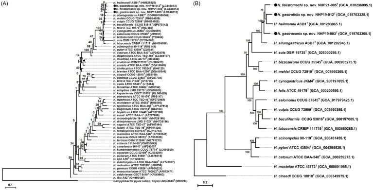
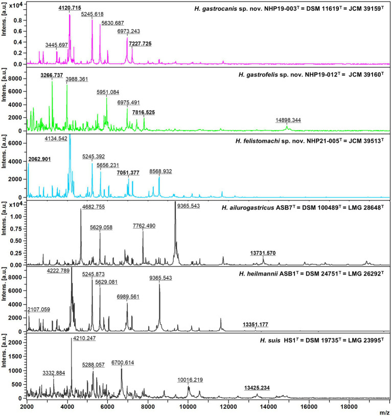
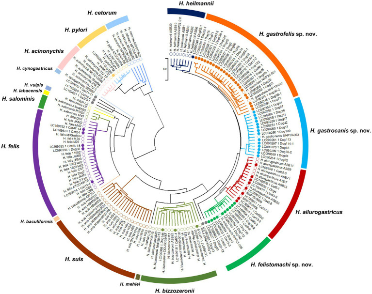
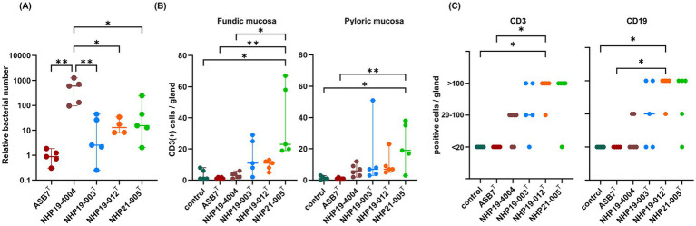
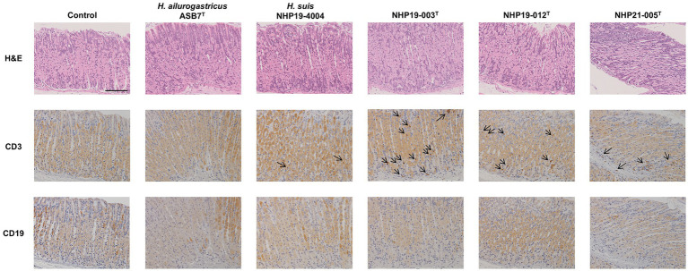
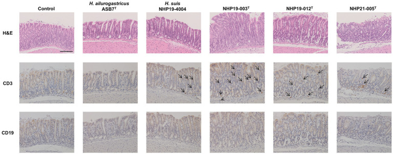
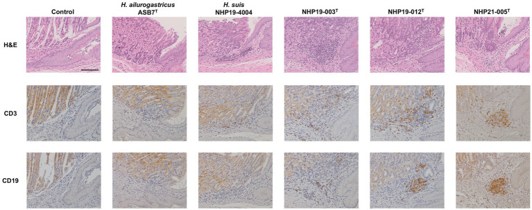
Helicobacter species infecting the stomachs of dogs and cats are potentially pathogenic and have been isolated from patients with gastric diseases. In the present study conducted in Japan, among the nine Helicobacter strains that we isolated from dogs and cats, NHP19-003T from a dog, and strains NHP19-012T and NHP21-005T from cats were identified to be the strains most closely related to Helicobacter heilmannii ASB1T based on a 16S rRNA comparison (98.7-99.2% similarity with H. heilmannii ASB1T). However, none of their whole genomes showed more than average nucleotide identity (ANI) threshold value (95-96%) to any Helicobacter species (85.1, 86.7, and 86.6% ANI, respectively, with H. heilmannii ASB1T), including when compared to each other. Furthermore, NHP19-003T, NHP19-012T, and NHP21-005T exhibited protein profiles different from known gastric Helicobacter species, as revealed by MALDI-TOF MS, indicating that they are novel Helicobacter species. We, thus, propose these novel Helicobacter species as follows: Helicobacter gastrocanis sp. nov. (type strain NHP19-003T [=JCM 39159T = DSM 111619T]), Helicobacter gastrofelis sp. nov. (type strain NHP19-012T [=JCM 39160T]) and Helicobacter felistomachi sp. nov. (type strain NHP21-005T [=JCM 39513T]). These novel strains have respective GC content values of 48.3, 46.9, and 47.1%. Phylogenetic analysis based on ureAB gene sequences obtained from gastric specimens from 47 dogs and 24 cats in Japan revealed that 29.8% of dogs were infected with H. gastrocanis, while H. gastrofelis infected 44.7% of dogs and 12.5% of cats. Additionally, 10.6% of dogs and 20.8% of cats were infected with H. felistomachi. Animal experiments have confirmed that these three novel species elicit gastric inflammatory responses. This study findings reveal the prevalence of novel gastric Helicobacter species in dogs and cats in Japan and their pathogenicity.

Keywords: cats; dogs; gastric Helicobacter species; gastric disease; novel species.


Conflict of interest statement

The authors declare that the research was conducted in the absence of any commercial or financial relationships that could be construed as a potential conflict of interest.

Figures

Figure 1
Figure 1
Average nucleotide identity (ANI) of the nine strains analyzed in this study compared to other Helicobacter species. A heatmap and hierarchical clustering are shown. Pairs of genomes with ANI > 95% can be considered as from the same species. The nine strains shown in bold are those isolated in this study.
Figure 2
Figure 2
Endoscopic and histological images from NHP19-003T–infected dog, NHP19-012T–infected cat, and NHP21-005T–infected cat. Representative endoscopic images NHP19-003T–infected dog suffering from protein-losing enteropathy (A), NHP19-012T–infected cat suffering from large-cell lymphoma (B), and NHP21-005T–infected cat suffering from gastric lymphoma (C). Giemsa staining of gastric mucosa of infected dog (D) and infected cat (E,F). Giemsa staining showed the presence of corkscrew-like morphology of the bacteria (arrows). Scanning electron micrograph of strains NHP19-003T (G), NHP19-012T (H), and NHP21-005T (I). Electron microscopy showed the spiral bacilli with a characteristic twisted structure with multiple flagella at both ends.
Figure 3
Figure 3
A circular genomic map of NHP19-003T, NHP19-012T, and NHP21-005T, comparing a circular graphical display of the distribution of genome information. The gene maps of (A) Helicobacter gastrocanis sp. nov. NHP19-003T; (B) Helicobacter gastrofelis sp. nov. NHP19-012T; and (C) Helicobacter felistomachi sp. nov. NHP21-005T. VFG, virulence factor gene; CDS, coding sequence.
Figure 4
Figure 4
Phylogenetic tree, conserved regions, and domains in VacA of H. pylori ATCC43504T and VacA-like proteins of H. heilmannii ASB1T, H. ailurogastricus ASB7T, H. suis HS1T, NHP19-003T, NHP19-012T, NHP21-005T, and H. pylori ATCC43504T. The amino acid sequences of VacA and VacA-like proteins were aligned by MAFFT version 7.49, and phylogenetic tree was constructed using RAxML-NG version 1.1.0 with an LG + G4 model and 1,000 bootstrap replicates. Numbers indicate bootstrap percentages, and the scale bar indicates the number of base substitutions per site. Conserved domains were identified using the NCBI platform’s CD-search tool. The conserved regions and identified domains were visualized using CLC Genomics Workbench Version 22.0.2.
Figure 5
Figure 5
Phylogenetic trees showing the relationship of NHP19-003T, NHP19-012T and NHP21-005T to their closely related species. (A) Is created from 16S rRNA gene sequences of Helicobacter species including NHP19-003T, NHP19-012T, NHP21-005T and 54 Helicobacter species. Campylobacter jejuni LMG 8841T was added as an out group. The sequences were aligned by MAFFT version 7.49 and the phylogenetic tree was constructed using RAxML-NG version 1.1.0 with a GTR + G + I model and 1,000 bootstrap replicates. Numbers indicate bootstrap percentages, and the scale bar indicates the number of base substitutions per site. Circles indicated Helicobacter sp. strain NHP19-003T, NHP19-012T, and NHP21-005T, respectively. (B) Is generated from 230 core genes of gastric Helicobacter species. Core genes alignments were obtained from Roary and aligned sequences were used for the phylogenetic tree reconstruction using RAxML-NG version 1.1.0 with a GTR + G + I model and 1,000 bootstrap replicates. H. cinaedi was included as an out group. Numbers and the scale bar indicate bootstrap percentages and substitutions per nucleotide position, respectively. Circles indicate Helicobacter sp. strain NHP19-003T, NHP19-012T, and NHP21-005T, respectively.
Figure 6
Figure 6
Comparative MALDI-TOF spectra profiles of type strains of novel species and related Helicobacter species strains obtained with flexAnalysis software. The baseline was subtracted. The y-axis shows the relative intensities of the ions, and the x-axis shows ion masses (Da). The peak at m/z approximately 13,000 pertaining to three other gastric Helicobacter species, H. ailurogastricus, H. heilmannii, and H. suis was not detected in NHP19-003T, NHP19-012T, and NHP21-005T.
Figure 7
Figure 7
Phylogenetic tree generated from ureAB gene sequences of gastric Helicobacter species. The ureAB sequences shown as the GenBank no. (LCXXXXXX) from cats (n = 24) and dogs (n = 47) in Japan analyzed in previous studies (13, 14). The ureAB sequences obtained from nine strains analyzed in this study (NHP19-003T, NHP19-012T, NHP21-005T, NHP22-001, NHP19-002, NHP19-009, NHP20-010, NHP20-013, and NHP21-011), and 88 reference strains of gastric Helicobacter species strains whose genomic sequences were available from NCBI were added to the analysis. The accession numbers of the ureAB sequences used in the analysis are summarized in Supplementary Table S4. The sequences were aligned by MAFFT version 7.49 and the tree was constructed using RAxML-NG version 1.1.0 with a GTR + G + I model and 1,000 bootstrap replicates. Numbers indicate bootstrap percentages, and the scale bar indicates the number of base substitutions per site. Open circles are 88 reference strains and nine strains analyzed in this study, and closed circles are 71 samples obtained from cats and dogs in Japan in previous studies (13, 14). Light blue, orange and green open circles indicate Helicobacter sp. strain NHP19-003T, NHP19-012T, NHP22-001 and NHP21-005T, respectively. The ureAB sequences in light blue, orange, and green subtrees were predicted to belong to the same species as Helicobacter sp. strain NHP19-003T, NHP19-012T, and NHP21-005T, respectively.
Figure 8
Figure 8
Effect of novel Helicobacter species infection in mouse stomachs. (A) Relative bacterial number of Helicobacter species in the mouse stomach. Bars indicate medians with 95% confidence interval. (B) Number of CD3(+) cells per glands in fundic and pyloric mucosa. (C) Number of CD3(+) cells and CD19(+) cells in the forestomach-glandular border. *P < 0.05, **P < 0.01.
Figure 9
Figure 9
Effect of novel Helicobacter species infection in fundic mucosa of mouse stomachs. H&E, CD3, and CD19 staining in fundic mucosa of stomach sections of Helicobacter species-infected and control mice. CD3(+) lymphocytes (arrows) were observed in NHP19-003T, NHP19-012T, and NHP21-005T–infected mice, while most lymphocytes were mostly negative for CD19. Bar indicates 100 μm.
Figure 10
Figure 10
Effect of novel Helicobacter species infection in pyloric mucosa of mouse stomachs. H&E, CD3, and CD19 staining in pyloric (E) mucosa of stomach sections of Helicobacter species-infected and control mice. CD3(+) lymphocytes (arrows) were observed in NHP19-003T, NHP19-012T, and NHP21-005T–infected mice, while most lymphocytes were mostly negative for CD19. Bar indicates 100 μm.
Figure 11
Figure 11
Effect of novel Helicobacter species infection in forestomach-glandular border of 689 mouse stomachs. H&E, CD3, and CD19 staining in the forestomach-glandular border of stomach sections from Helicobacter species-infected and control mice. Bar indicates 100 mm.

Similar articles

Cited by

References

    1. Amorim I., Smet A., Alves O., Teixeira S., Saraiva A. L., Taulescu M., et al. . (2015). Presence and significance of Helicobacter spp. in the gastric mucosa of Portuguese dogs. Gut Pathogens 7:12. doi: 10.1186/s13099-015-0057-1, PMID: - DOI - PMC - PubMed
    1. Baele M., Decostere A., Vandamme P., Van den Bulck K., Gruntar I., Mehle J., et al. . (2008). Helicobacter baculiformis sp. nov., isolated from feline stomach mucosa. Int. J. Syst. Evol. Microbiol. 58, 357–364. doi: 10.1099/ijs.0.65152-0, PMID: - DOI - PubMed
    1. Dewhirst F. E., Shen Z., Scimeca M. S., Stokes L. N., Boumenna T., Chen T., et al. . (2005). Discordant 16S and 23S rRNA gene phylogenies for the genus Helicobacter: implications for phylogenetic inference and systematics. J. Bacteriol. 187, 6106–6118. doi: 10.1128/jb.187.17.6106-6118.2005, PMID: - DOI - PMC - PubMed
    1. Frith M. C., Wan R., Horton P. (2010). Incorporating sequence quality data into alignment improves DNA read mapping. Nucleic Acids Res. 38:e100. doi: 10.1093/nar/gkq010, PMID: - DOI - PMC - PubMed
    1. Goodwin C. S., Armstrong J. A., Chilvers T., Peters M., Collins M. D., Sly L., et al. . (1989). Transfer of Campylobacter pylori and Campylobacter mustelae to Helicobacter gen. Nov. as Helicobacter pylori comb. nov. and Helicobacter mustelae comb, nov., respectively. Int. J. Syst. Evol. Microbiol. 39, 397–405. doi: 10.1099/00207713-39-4-397, PMID: - DOI - PubMed

LinkOut - more resources