. 2025 Jan 7:10:100268.
doi: 10.1016/j.jtauto.2025.100268. eCollection 2025 Jun.

Across ancestries, HLA-B∗08:01∼DRB1∗03:01 (DR3) and HLA-DQA∗01:02 (DR2) increase the risk to develop juvenile-onset systemic lupus erythematosus through low complement C4 levels

Collaborators, Affiliations

Across ancestries, HLA-B∗08:01∼DRB1∗03:01 (DR3) and HLA-DQA∗01:02 (DR2) increase the risk to develop juvenile-onset systemic lupus erythematosus through low complement C4 levels

Yves Renaudineau et al. J Transl Autoimmun. .

Abstract

Objective: Systemic lupus erythematosus (SLE) is a systemic autoimmune/inflammatory disease with a strong genetic component. Genetic burden is higher in children when compared to patients with adult-onset SLE, contributing to earlier disease expression and more severe phenotypes. The human leukocyte antigen (HLA) cluster on chromosome 6p21.3 is among the most variable genomic regions, representing a major risk-factor for SLE in adults. Its impact on juvenile-onset (j)SLE remains largely unstudied.

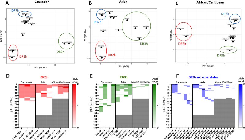
Methods: High-resolution sequencing of HLA class I (A, B, C), class II (DRB1, DQA1, DQB1) and class III (complement C2) was undertaken in the multi-ancestral UK JSLE Cohort including participants of Caucasian (n = 151, 48.8 %), Asian (n = 108, 35.0 %) and African/Caribbean (n = 50, 16.2 %) descent. Considering ancestral variation, clinical associations were tested at the level of alleles (2-field resolution), associated HLA protein sequences (antigen binding domains, 4-field resolution), and extended haplotypes (DRh).

Results: Although important ancestral recombination was reported for HLA-DR2 and -DR3 haplotypes, risk associated with jSLE was conserved at related alleles (DR2h: DRB1∗15:01, DQA∗01:02, DQB1∗06:02; DR3h: C∗07:02 [Asian], B∗08:01, C2 rs9332730 [Asian], DRB1∗03:01). HLA-DR7 haplotypes (DRB1∗07:01, OR = 0.44, 95 % CI:0.27-0.72, p = 0.0004; DQA1∗02:01, OR = 0.34, 95 % CI:0.21-0.56, p = 1.8 × 10⁻⁶) protect Asians from jSLE development. Among 23 clinical variables recorded, the main association was found between low levels of complement C4 in Caucasian carriers of HLA-DR3h. This was not the case in Asians due to recombination with HLA-C∗07:02 and integration of the C2 rs9332730 minor allele. Low C4 serum levels associated with HLA-DQA1∗01:02 (DR2h) in Caucasians after excluding HLA-DR3h carriers from the analysis. An association between low white blood cell counts and HLA-A∗03:01P was observed across ancestries.

Conclusion: Genetic variation in the HLA cluster associates with organ domain involvement (hematological) and complement levels in jSLE. Lupus-associated HLA haplotypes vary between ancestral groups, underscoring the importance of multi-ancestral approaches to genetic studies in SLE and other autoimmune/inflammatory diseases.

Keywords: C4; Complement; Disease activity; Genetic; HLA; Haplotype; Juvenile-onset; Lupus; SLE.


Conflict of interest statement

The authors report no conflict of interest.

Figures

Fig. 1
Fig. 1
Ancestry-differential HLA allele diversity in jSLE. A: Manhattan plot according to ancestral groups: Caucasians in blue, Asians in red, African/Caribbean descendants in green. Ancestry-related controls were obtained from several databases (Materials and Methods). HLA-class I, HLA-class III (C2 gene), and HLA-class II regions are represented on the horizontal axis with – Log10 (p values) on the vertical axis. The statistical threshold was fixed at pFDR = 0.001; and the p = 0.01 cut-off is presented (doted lines). B-D: Odds ratios of HLA alleles significantly associated with jSLE across ethnicities. Significant risk and protective factors are displayed in colour (pFDR<0.001).
Fig. 2
Fig. 2
HLA-DR2(h), -DR3h, and -DR7h haplotypes differentially associate with jSLE across ancestral groups. A-C: Principal component analysis (PCA) based on 14 risk/protective alleles identified in jSLE patients (Fig. 1) of Caucasian (A), Asian (B) or African/Caribbean ancestry (C). D: HLA-DR2h, E: HLA-DR3h and F: HLA-DR7h and other alleles according to the ancestry group.
Fig. 3
Fig. 3
jSLE-associated HLA quantitative trait loci (QTL). A:Manhattan plot of HLA alleles (x A∗, x C∗, B∗, x C2-factor B, x DRB1∗, x DQA1∗, and xDQB1∗) with a frequency ≥0.05 and their association with age at diagnosis (years), disease activity (pBILAG 2004, SLEDAI-2K), disease activity (pBILAG 2004 organ domains), and clinical features.B:Concordance with HLA protein sequence.C:Concordance with haplotypes DR2(h), DR3h, DR7h and ancestral variants. On the y axis,–Log10(p values) are presented, assessing concordance between allele/protein number (0, 1, and 2) or haplotype (DR3h/DR7h: 0, 2, 3, and 4 alleles; DR2h: 0, 2, 3, 4, 5, and 6 alleles), and the quantitative trait using the Spearman's correlation test. Alleles with at least one pFDR value ≤ 0.001 are indicated as full circles and those with a least one p value 0.01≤p < 0.001 are reported as an empty circles (A and B). Dark vertical lines represent p value thresholds at -LOG10 = 2 (p = 0.01) and -LOG10 = 3 (p = 0.001). C3: complement factor 3; C4: complement factor 4; anti-dsDNA: anti-double stranded DNA antibodies; anti-Sm: anti-Smith antibodies; anti-RNP: anti-ribonucleopeptide antibodies; Hb: hemoglobin; WBC: white blood cell count; Plt: platelet count; ESR: erythrocyte sedimentation rate.
Fig. 4
Fig. 4
jSLE-associated HLA quantitative trait loci after excluding individuals harboring the ancestral DR3 haplotype (B∗08:01-X-DRB1∗03:01). A: Manhattan plot of HLA alleles (x A∗, x C∗, B∗, x C2-factor B, x DRB1∗, x DQA1∗, and xDQB1∗) and their association with jSLE features. B: Concordance with HLA protein sequence. C: Concordance with haplotypes HLA-DR2, -DR3, -DR7 and ancestral variants. On the x axis, –Log10(p values) are presented, assessing the concordance between allele/protein number (0, 1, and 2) or haplotype (HLA-DR3/DR7h: 0, 2, 3, and 4 alleles; HLA-DR2h: 0, 2, 3, 4, 5, and 6 alleles) and quantitative traits using the Spearman's correlation test. Alleles with at least one pFDR value ≤ 0.001 are indicated as full circles and those with a least one p value 0.01≤p < 0.001 are reported as an empty circles (A, B). Dark vertical lines represent p value thresholds at -LOG10 = 2 (p = 0.01) and -LOG10 = 3 (p = 0.001). C3: complement factor 3; C4: complement factor 4; anti-dsDNA: anti-double stranded DNA antibodies; anti-Sm: anti-Smith antibodies; anti-RNP: anti-ribonucleopeptide antibodies; Hb: hemoglobin; WBC: white blood cell count; Plt: platelet count; ESR: erythrocyte sedimentation rate.

References

    1. Charras A., Smith E., Hedrich C.M. Systemic lupus erythematosus in children and young people. Curr. Rheumatol. Rep. 2021;23(3):20. - PMC - PubMed
    1. Tsokos G.C. The immunology of systemic lupus erythematosus. Nat. Immunol. 2024;25(8):1332–1343. - PubMed
    1. Massias J.S., Smith E.M.D., Al-Abadi E., Armon K., Bailey K., Ciurtin C., et al. Clinical and laboratory characteristics in juvenile-onset systemic lupus erythematosus across age groups. Lupus. 2020;29(5):474–481. - PMC - PubMed
    1. Ambrose N., Morgan T.A., Galloway J., Ionnoau Y., Beresford M.W., Isenberg D.A., et al. Differences in disease phenotype and severity in SLE across age groups. Lupus. 2016;25(14):1542–1550. - PMC - PubMed
    1. Massias J.S., Smith E.M., Al-Abadi E., Armon K., Bailey K., Ciurtin C., et al. Clinical and laboratory phenotypes in juvenile-onset Systemic Lupus Erythematosus across ethnicities in the UK. Lupus. 2021;30(4):597–607. - PMC - PubMed

LinkOut - more resources