

Surface functionalization of microscaffolds produced by high-resolution 3D printing: A new layer of freedom
- PMID: 39896295
- PMCID: PMC11783114
- DOI: 10.1016/j.mtbio.2025.101452
Surface functionalization of microscaffolds produced by high-resolution 3D printing: A new layer of freedom
Abstract
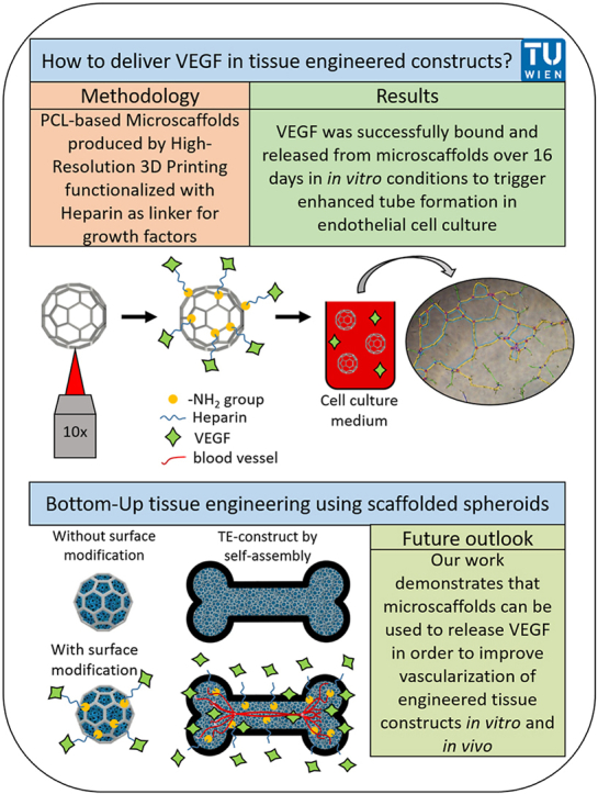
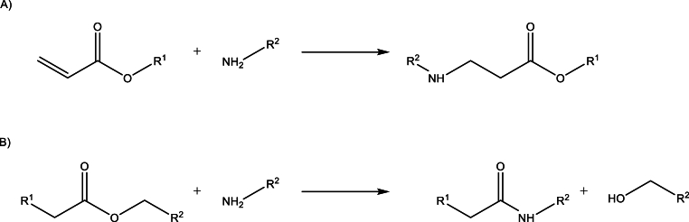
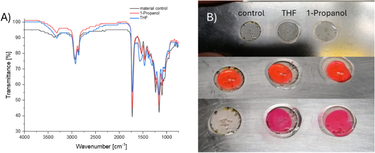
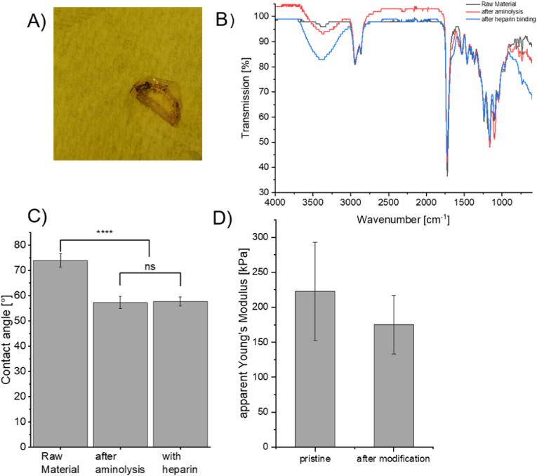
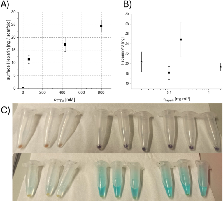
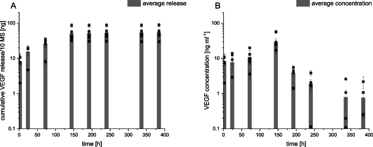
Scaffolded-spheroids represent novel building blocks for bottom-up tissue assembly, allowing to produce constructs with high initial cell density. Previously, we demonstrated the successful differentiation of such building blocks, produced from immortalized human adipose-derived stem cells, towards different phenotypes, and the possibility of creating macro-sized tissue-like constructs in vitro. The culture of cells in vitro depends on the supply of various nutrients and biomolecules, such as growth factors, usually supplemented in the culture medium. Another means for growth factor delivery (in vitro and in vivo) is the release from the scaffold to alter the biological response of surrounding cells (e.g. by release of VEGF).1 As a proof of concept for this approach, we sought to biofunctionalize the surface of the microscaffolds with heparin as a "universal linker" that would allow binding a variety of growth factors/biomolecules. An aminolysis step in an organic solvent made it possible to generate a hydrophilic and charged surface. The backbone of the amine, as well as reaction conditions, led to an adjustable surface modification. The amount of heparin on the surface was increased with an ethylene glycol-based diamine backbone and varied between 8 and 40 ng per microscaffold. Choosing a suitable linker allows easy adjustment of the loading of VEGF and other heparin-binding proteins. Initial results indicated that up to 5 ng VEGF could be loaded per microscaffold, generating a steady VEGF release for 16 days. We report an easy-to-perform, scalable surface modification approach of polyester-based resin that leads to adjustable surface concentrations of heparin. The successful surface aminolysis opens the route to various modifications and broadens the spectrum of biomolecules which can be delivered.
Keywords: Growth factors; High-resolution 3D printing; Microscaffolds; Scaffolded spheroids; Surface modification; Tissue engineering; VEGF.
© 2025 The Authors.
Conflict of interest statement
The authors declare the following financial interests/personal relationships which may be considered as potential competing interests:Aleksandr Ovsianikov is co-founder of UpNano GmbH, a TU Wien spin-off active in the area of two-photon polymerization. His current relationship with UpNano includes: consulting, advisory and equity. The rest of the authors declare that they have no known competing financial interests or personal relationships that could have appeared to influence the work reported in this manuscript.
Figures
Similar articles
-
Hybrid spheroid microscaffolds as modular tissue units to build macro-tissue assemblies for tissue engineering.Acta Biomater. 2023 Jul 15;165:72-85. doi: 10.1016/j.actbio.2022.03.010. Epub 2022 Mar 12. Acta Biomater. 2023. PMID: 35288312
-
Scaffolded spheroids as building blocks for bottom-up cartilage tissue engineering show enhanced bioassembly dynamics.Acta Biomater. 2024 Jan 15;174:163-176. doi: 10.1016/j.actbio.2023.12.001. Epub 2023 Dec 7. Acta Biomater. 2024. PMID: 38065247
-
Optical µ-Printing of Cellular-Scale Microscaffold Arrays for 3D Cell Culture.Sci Rep. 2017 Aug 21;7(1):8880. doi: 10.1038/s41598-017-08598-3. Sci Rep. 2017. PMID: 28827528 Free PMC article.
-
Adult Stem Cells Spheroids to Optimize Cell Colonization in Scaffolds for Cartilage and Bone Tissue Engineering.Int J Mol Sci. 2018 Apr 25;19(5):1285. doi: 10.3390/ijms19051285. Int J Mol Sci. 2018. PMID: 29693604 Free PMC article. Review.
-
Cartilage and bone tissue engineering using adipose stromal/stem cells spheroids as building blocks.World J Stem Cells. 2020 Feb 26;12(2):110-122. doi: 10.4252/wjsc.v12.i2.110. World J Stem Cells. 2020. PMID: 32184936 Free PMC article. Review.
References
-
- Ovsianikov A., Khademhosseini A., Mironov V. The synergy of scaffold-based and scaffold-free tissue engineering strategies. Trends Biotechnol. 2018;36(4):348–357. - PubMed
-
- Moldovan N.I., Hibino N., Nakayama K. Principles of the Kenzan Method for Robotic Cell Spheroid-Based Three-Dimensional Bioprinting. Tissue Eng. B Rev. 2017;23(3):237–244. - PubMed
-
- Murata D., Arai K., Nakayama K. Scaffold-free bio-3D printing using spheroids as “bio-inks” for tissue (Re-)Construction and drug response tests. Adv. Healthcare Mater. 2020;9(15) - PubMed
LinkOut - more resources
Full Text Sources
Research Materials
