[Preprint]. 2025 Jan 23:2025.01.23.634564.
doi: 10.1101/2025.01.23.634564.

Pancreatic injury induces β-cell regeneration in axolotl

Affiliations

Pancreatic injury induces β-cell regeneration in axolotl

Connor J Powell et al. bioRxiv. .

Update in

  • Pancreatic injury induces β-cell regeneration in axolotl.
    Powell CJ, Singer HD, Juarez AR, Kim RT, Kim E, Payzin-Dogru D, Savage AM, Lopez NJ, Thornton K, Blair SJ, Abouelela A, Dittrich A, Akeson SG, Jain M, Whited JL. Powell CJ, et al. Dev Dyn. 2025 Jul 18:10.1002/dvdy.70060. doi: 10.1002/dvdy.70060. Online ahead of print. Dev Dyn. 2025. PMID: 40679186

Abstract

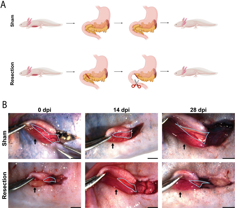
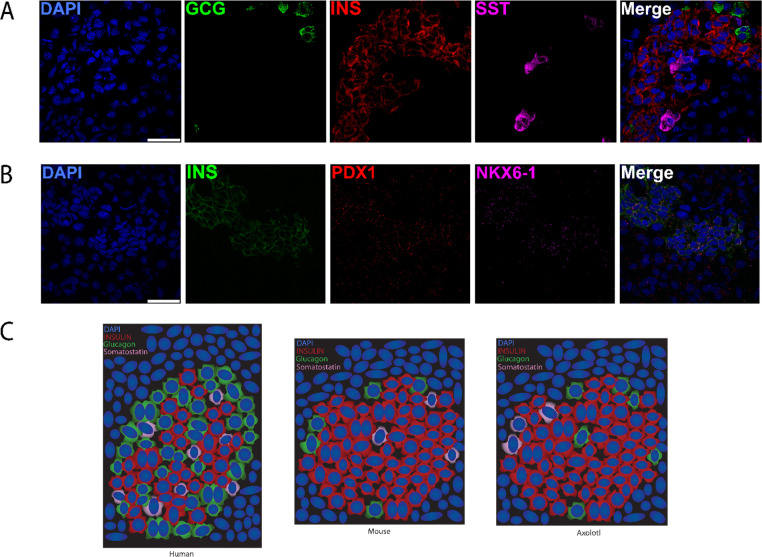
Background: Diabetes is a condition characterized by a loss of pancreatic β-cell function which results in the dysregulation of insulin homeostasis. Using a partial pancreatectomy model in axolotl, we aimed to observe the pancreatic response to injury.

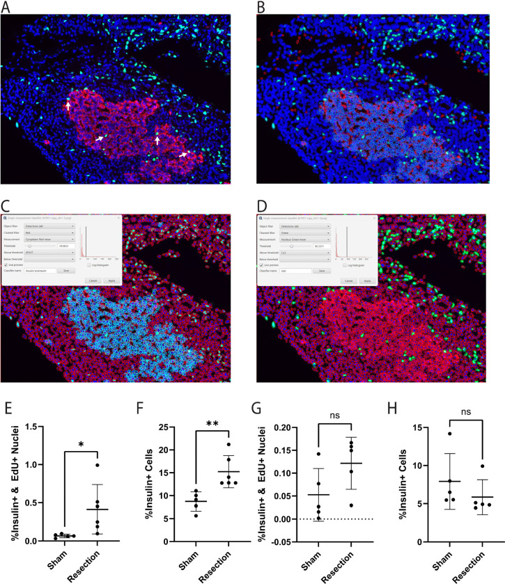
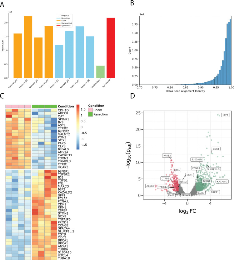
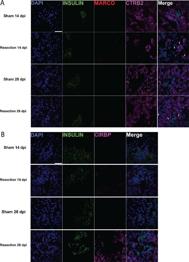
Results: Here we show a comprehensive histological assessment of pancreatic islets in axolotl. Following pancreatic injury, no apparent blastemal structure was observed. We found a significant, organ-wide increase in cellular proliferation post-resection in the pancreas compared to sham-operated controls. This proliferative response was most robust at the site of injury. We found that β-cells actively contributed to the increased rates of proliferation upon injury. β-cell proliferation manifested in increased β-cell mass in injured tissue at two weeks post injury. At four weeks post injury, we found organ-wide proliferation to be extinguished while proliferation at the injury site persisted, corresponding to pancreatic tissue recovery. Similarly, total β-cell mass was comparable to sham after four weeks.

Conclusions: Our findings suggest a non-blastema-mediated regeneration process takes place in the pancreas, by which pancreatic resection induces whole-organ β-cell proliferation without the formation of a blastemal structure. This process is analogous to other models of compensatory growth in axolotl, including liver regeneration.

Keywords: compensatory growth; diabetes; insulin; pancreatectomy; proliferation; resection.


Conflict of interest statement

Declaration of Interests JLW is a co-founder of Matice Biosciences. Other authors declare no competing interests.

Figures

Figure 1.
Figure 1.. Pancreatic resection surgery model.
A. Cartoon depicting sham and pancreatic resection surgery. B. Morphology of sham and pancreatic resection surgery at 0, 14 and 28 dpi. The pancreas is outlined in blue while black arrows point to the duodenum. 0 dpi image of the resection condition was taken immediately following 20% removal of pancreas by length. The majority of the pancreas extends into the abdominal cavity where it connects to the liver. In each image, the orientation from left to right follows a rostral to caudal direction. Black scale bars in the bottom right corner of each image are 2 mm.
Figure 2.
Figure 2.. Pancreatic resection initially provokes substantial proliferative responses throughout the entire organ, but cell proliferation becomes restricted local to the site of injury later in the process.
A. EdU and insulin (IHC) stain of pancreas samples at 14 dpi. B. Organ-wide proliferation is increased in resection samples at 14 dpi (p=0.0162) (sham n=5, resection n=6). C. Proliferation local to the site of injury is higher within resection samples at 14 dpi (p=0.0032). D. Proliferation local to the site of injury is increased at 14 dpi (p=0.0012). E. EdU and insulin stain of pancreas samples at 28 dpi (sham n=5, resection n=5). F. Organ-wide proliferation in resection samples is non-significant in comparison to sham at 28 dpi (p=0.8459). G. Proliferation local to the site of injury is higher within resection samples at 28 dpi (p=0.0085). H. Proliferation local to the site of injury is increased at 28 dpi (p=0.0079). I. Cartoon representation of how the area local to the site of injury was chosen. J. Organ-wide proliferation is decreased between resection samples from 14 to 28 dpi (p=0.0009). K. Local proliferation is decreased between resection samples from 14 to 28 dpi (p=0.0005). Scale bars are 100 μm.
Figure 3.
Figure 3.. β–cell proliferation is observed at 14 days post injury but subsides by 28 days.
A. Representative image of a coronal section of regenerating pancreatic tissue at 14 dpi. White arrows point to proliferating β–cells. B. Visualization of image quantification with QuPath Software. Cells were categorized by expression of insulin/ proinsulin antibody and EdU stain, then counted for downstream statistical analysis. C. Settings used to identify insulin/ proinsulin positive cells. Light blue outlines represent insulin/ proinsulin positive cells. Red outlines represent cells negative for insulin/ proinsulin. D. Settings used to identify EdU positive cells. Green outlines represent EdU positive cells. Red outlines represent EdU negative cells. E. A significant increase in cells positive for both EdU and insulin antibody was observed at 14 dpi (p = 0.0463) (sham n=5, resection n=6). F. A significant increase in insulin positive cells was observed at 14 dpi (p = 0.005) (Sham n=5, Resection n=6). G. No significant difference in cells positive for both EdU and insulin antibody was observed at 28 dpi (p = 0.0936) (Sham n=5, Resection n=5). H. No significant difference in insulin positive cells was observed at 28 dpi (p = 0.3181) (Sham n=5, Resection n=5).
Figure 4.
Figure 4.. Nanopore sequencing data reveals transcriptomic changes in response to injury.
A. Bar plot detailing reads per sample after demultiplexing. Unclassified reads and reads with a qscore less than 8 were filtered out. B. Histogram detailing aligned read identity vs total read counts. C. Heatmap of differentially expressed genes at 14 dpi D. Volcano plot of differentially expressed genes at 14 dpi.
Figure 5:
Figure 5:. Genes previously implicated in regeneration exhibit upregulation in axolotl pancreatic injury model.
A. HCR-FISH showing differential expression of insulin and Marco transcripts. Ctrb2 was not found to be differentially expressed; however, it provides structural context for exocrine tissue. Marco+ cells shown with arrows. B. HCR-FISH showing differential expression of insulin and Cirbp transcripts. Scale bars are 50 μm.
Figure 6.
Figure 6.. Transcript level pancreatic islet morphology in axolotl
A. HCR-FISH of glucagon (Gcg), insulin (Ins), and somatostatin (Sst) transcripts. B. HCR-FISH of insulin (Ins), Pdx1, and Nkx6–1 transcripts. C. Cartoon representation comparing axolotl islets to mice and humans. Mouse and human islet cartoons based on data from Abdulreda et al. Scale bars are 50 μm.

References

    1. Vos T., et al. , 2020. Global burden of 369 diseases and injuries in 204 countries and territories, 1990–2019: a systematic analysis for the Global Burden of Disease Study 2019. The Lancet 396, 1204–1222.. 10.1016/s0140-6736(20)30925-9 - DOI - PMC - PubMed
    1. Roger V.L., 2021. Epidemiology of Heart Failure. Circulation Research 128, 1421–1434.. 10.1161/circresaha.121.318172 - DOI - PubMed
    1. Cheng N.-C., Tai H.-C., Chang S.-C., Chang C.-H., Lai H.-S., 2015. Necrotizing fasciitis in patients with diabetes mellitus: clinical characteristics and risk factors for mortality. BMC Infectious Diseases 15.. 10.1186/s12879-015-1144-0 - DOI - PMC - PubMed
    1. Zhou Q., Melton D.A., 2018. Pancreas regeneration. Nature 557, 351–358.. 10.1038/s41586-018-0088-0 - DOI - PMC - PubMed
    1. Vegas A.J., Veiseh O., Gürtler M., Millman J.R., Pagliuca F.W., Bader A.R., Doloff J.C., Li J., Chen M., Olejnik K., Tam H.H., Jhunjhunwala S., Langan E., Aresta-Dasilva S., Gandham S., Mcgarrigle J.J., Bochenek M.A., Hollister-Lock J., Oberholzer J., Greiner D.L., Weir G.C., Melton D.A., Langer R., Anderson D.G., 2016. Long-term glycemic control using polymer-encapsulated human stem cell–derived beta cells in immune-competent mice. Nature Medicine 22, 306–311.. 10.1038/nm.4030 - DOI - PMC - PubMed

Publication types

LinkOut - more resources