[Preprint]. 2025 Jan 26:2025.01.24.634764.
doi: 10.1101/2025.01.24.634764.

Phosphatidylserine (PS)-targeting chimeric Interferon (IFN) fusion proteins for anti-tumor applications

Affiliations

Phosphatidylserine (PS)-targeting chimeric Interferon (IFN) fusion proteins for anti-tumor applications

Varsha Gadiyar et al. bioRxiv. .

Abstract

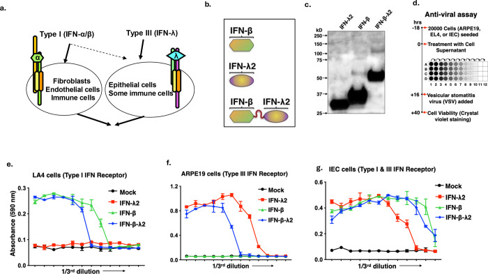
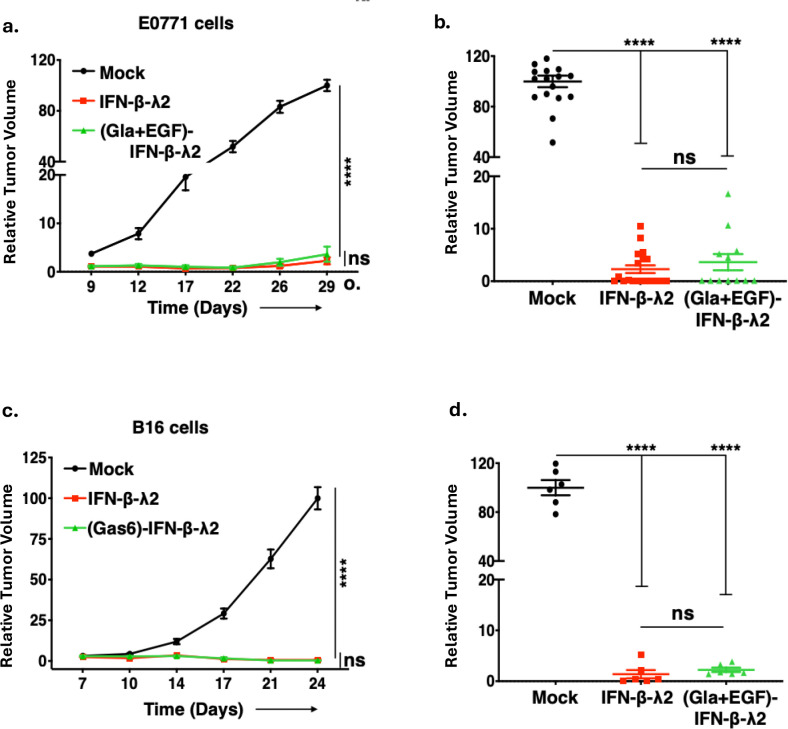
In viable healthy cells, membrane phospholipids are asymmetrically distributed across the lipid bilayer, whereby the anionic phospholipid phosphatidylserine is virtually all distributed on the inner leaflet of the plasma membrane. During apoptosis, phospholipid asymmetry collapses and PS is externalized to the external leaflet where it serves as an "eat-me" signal for efferocytosis, the process whereby dying cells are engulfed and degraded by phagocytes. PS is also externalized on viable activated tumor endothelial cells, stromal cells and cancer cells in the tumor microenvironment reflecting a pathophysiological state of solid cancers that function to suppress host anti-tumor immunity. Several strategies have been envisioned to target dysregulated PS in the tumor microenvironment including PS binding proteins such as Annexin V and PS-targeting monoclonal antibodies (Bavituximab) with promising preclinical results. Here, in an attempt to enhance the efficacy of PS-targeting therapeutics, we have generated a series of recombinant chimeric fusion proteins that fuse type I and type III IFNs (IFN-β-IFN-λ) into a single polypeptide chain separated by a short linker. The IFN-β-IFN-λ fusion proteins retain functions of both type I and type III IFNs but show combined effects to improve biological function as well as enhance anti-tumor activities. To localize IFNs to sites of externalized PS, we next fused the IFN-β-IFN-λ chimeric protein to the PS-targeting gamma-carboxyglutamic acid-rich (Gla) domain of Growth Arrest Specific factor 6 (Gas-6), rendering these IFN biologics as PS targeting modalities. Gas6-IFN-β-IFN-λ proteins selectively bind PS as evident by solid-phase ELISA assays as well as bind PS-positive cells, including apoptotic cells and cells that express CDC50 subunit mutant of the ATP11C flippase. In vivo, Gas6-IFN-β-IFN-λ retain strong anti-tumor activities in a syngeneic model when expressed ectopically in a E0771 breast cancer model and B16-F10 melanoma models. Collectively, we report on the generation and utility of a series of novel in class IFN fusion proteins that target the immune stimulatory features of IFNs to the PS externalization in the tumor microenvironment.

Keywords: Immune Escape; Immunogenic Biologicals; PS Targeting; PS receptors; Phosphatidylserine (PS); Type I Interferons; Type III Interferons.


Conflict of interest statement

Disclosure of potential conflicts of interest: SVK and RBB are cofounders of a biotechnology company called Targeron Therapuetics, LLC that aims to develop PS-targeting IFNs for immune-oncology applications,

Figures

Figure 1.
Figure 1.. Design, production and characterization of novel IFN fusion proteins.
(a) Action of IFNs are compartmentalized, where type I IFNS act on fibroblast, endothelial cells and most immune cells, whereas type III IFNs act mainly on epithelial cells. (b) The schematic illustration of IFN fusion proteins. (c) Immunoblot showing secretion of each protein at their expected molecular weight in the cell supernatant when probed using anti-his antibody. (d) The schematic illustration depicting antiviral assay strategy. The IFN receptor expressing cells were pretreated with HEK293T cell supernatant containing IFN, IFN fusion and Gas6-IFN fusion proteins. After 12 h of pretreatment, cells were treated for 24 h with vesicular stomatitis virus (VSV) and cell viability was determined by staining cells with 0.1% crystal violet solution followed by reading absorbance at 590nm. (e) The representative graphs showing the antiviral activity of IFNs in LA4, mouse lung adenoma cells (express type I IFN receptor) (f), ARPE19, the human retinal epithelium cells (respond only to mouse type III IFNs) (g), and mIEC, mouse intestine epithelium cells (express both type I and type III IFN receptors).
Figure 2.
Figure 2.. Anti-tumor effect of IFNs and IFN fusion proteins in the E0771 murine breast cancer tumor model.
(a) Antiviral assay (VSV) at 24 h pre-treatment with equivalent amounts of IFNs on IEC cells. Concentration at which IFN treatment showed 50% activity is plotted on Y axis (p=0.0025, n=3, multiple t-tests). (b)The schematic illustration showing transfection of E0771 or B16-F10 cells followed by antibiotic selection to create stable cell lines that constitutively secrete IFN fusion proteins. (c) The graphical illustration showing experimental outline of the E0771 murine breast cancer tumor model in C57BL/6 mice. 5 X 10⁴ E0771 cells, mock or constitutively secreting IFN- β, IFN-λ, or IFN- β -IFN-λ, or IFN- β and IFN-λ in 50:50 ratio were injected orthotopically into the mammary fat pad of female C57BL/6 mice. Tumor growth was monitored every 3–5 days for 4 weeks and tumor volumes are shown as relative mean (d, f) or individual (e, g) tumor volumes compared to the mean volume of mock-transfected tumors measured at the end point (day 29). (h) Mice injected with E0771 cells expressing IFN- β and IFN-λ in 50:50 showed ascites, whereas the mice injected with IFN- β -IFN-λ fusion did not show this phenotype. (i) Tumors expressing IFN- β -IFN-λ fusion protein, and livers from these mice show reduced anemia compared to the tumors expressing IFN- β and IFN-λ in 50:50.
Figure 3.
Figure 3.. Novel Gas6-IFN fusion proteins display both IFN activity and PS binding characteristics.
(a) Schematic showing the rationale for developing Gas6(Gla+EGF)-IFN-β-IFN-λ fusion proteins; Gla and EGF domains of Gas6 are required for PS binding, whereas, the LG domain activates immunosuppressive TAM receptor signaling. Truncating the Gas6, and replacing the LG domains with immunomodulatory IFN fusions will convert immunosuppressive signaling to pro-inflammatory signaling. (b) γ-carboxylated (PS binding) and non γ -carboxylated (non PS binding) variants of Gas6(Gla+EGF)-IFN-β-IFN-λ were produced by supplementing transfected HEK293T cell media with vitamin K or Warfarin respectively during protein production. (c) PS ELISA confirms that γ-carboxylated Gas6(Gla+EGF)-IFN-β-IFN-λ binds to PS whereas IFN-β-IFN-λ does not. (d) This is further confirmed by using CDC50AED29 cells, a mouse lymphoma cell line (W3) that harbors a mutation in CDC50, leading to the loss in PS-flippase function and constitutive externalization of PS on live cells. Gas6(Gla+EGF)-IFN-β-IFN-λ (VitK) binds to only CDC50AED29 (PS+) cells whereas Gas6(Gla+EGF)-IFN-β-IFN-λ (War) does not bind to either. (e) CHO IFN-λ reporter cells were treated with IFN-β-IFN-λ, Gas6(Gla+EGF)-IFN-β-IFN-λ (VitK) and Gas6(Gla+EGF)-IFN-β-IFN-λ (War) with CDC50AED29 cells (f) or apoptotic cells for 15 min, washed and centrifuged to remove unbound protein and incubated with reporter cells for 30 min. Immunoblot of pSTAT1 is used as a read out of IFN-λR activation in the reporter cells.
Figure 4.
Figure 4.. Anti-tumor effect of IFN-β-IFN-λ and Gas6-IFN-β-IFN-λ fusion molecules.
(a) Schematic diagram showing the different Gas6-IFN fusion proteins developed with just the Gla domain or the Gla and EGF domains of Gas6 with IFN-β, IFN-λ, or IFN-β-IFN-λ i.e. Gas6(Gla)-IFN-β, Gas6(Gla)-IFN-λ, Gas6(Gla)-IFN-β-IFN-λ, and Gas6(Gla+EGF)-IFN-β, Gas6(Gla+EGF)-IFN-λ, Gas6(Gla+EGF)-IFN-β-IFN-λ. (b) Immunoblot showing secretion of each protein at their expected molecular weight in the cell supernatant when probed using anti-His antibody and confirming the γ- carboxylation on the Gla domain using anti-Gla antibody (c). (d) Activation of Mertk-γ-R1 reporter cell line using Gas6 and PS shows that addition of PS in the form of apoptotic cells or PS liposomes shows hyperactivation of Mertk. Imaging flow cytometry shows distinct pattern of fusion protein binding to CDC50AED29 (PS+) cells. (e) Gas6-IFN-β-IFN-λ (VitK) shows clustered pattern of binding, whereas (f) IFN-β-IFN-λ2 shows a more diffuse pattern of staining. (g) shows enlarged images. (h) Quantification of the diffuse vs the clustering pattern shows more diffuse pattern with the IFN-β-IFN-λ2 versus, clustered pattern with Gas6-IFN-β-IFN-λ (VitK).
Figure 5:
Figure 5:. Novel fusion proteins show anti-tumor efficacy in E0771 and B16-F10 tumor models.
In vivo tumor growth curves of E0771 and B16-F10 tumor models. 5 X 10⁴ E0771 cells, mock transfected, or constitutively secreting either Gas6(Gla)-IFN-β-IFN-λ, Gas6(Gla+EGF)-IFN-λ2, Gas6(Gla+EGF)-IFN-β or Gas6(Gla+EGF)-IFN-β-IFN-λ, were injected orthotopically into the mammary fat pad of C57BL/6 mice and tumor growth determined by tumor volume measurements every 3–5 days for a period of 4 weeks. To compare anti-tumor effects of Gas6-IFN-β-IFN-λ fusion protein to the effects of Gas6-IFN-β and Gas6-IFN-λ (single IFN proteins) given in combination, C57BL/6 mice were implanted with 5 X 10⁴ E0771 cells producing Gas6(Gla+EGF)-IFN-β-IFN-λ cells or a 50:50 mixture of E0771 cells producing Gas6(Gla+EGF)-IFN-β and E0771 cells producing Gas6(Gla+EGF)-IFN-λ (total number of implanted cells 5 X 10⁴). The Gla+EGF domains and the fusion of IFNs β and λ have an additive effect, as shown by reduction in (a) tumor volume by days and (b) tumor volume at end point. Similarly, The Gas6(Gla+EGF)-IFN-β-IFN-λ fusion protein was much more potent in inhibiting tumor growth than the 50:50 combination of single IFNs as shown by relative tumor volume at various time points, and at end point (c,d).
Figure 6:
Figure 6:. Addition of the Gla and EGF domains do not interfere with anti-tumor activity.
To compare the effect of PS targeting on anti-tumor potency of IFN-β-IFN-λ fusion molecule, Gas6(Gla+EGF)-IFN-β-IFN-λ and IFN-β-IFN-λ were compared head to head in E0771 (a, b) and B16-F10 models (c, d). 5 X 10⁴ E0771 or 5 X 10⁴ B16 cells, mock-transfected or constitutively secreting either IFN-b-IFN-λ or Gas6(Gla+EGF)-IFN-β-IFN-λ, were injected either orthotopically into the mammary fat pad of female C57BL/6 mice for the E0771 model or into the right flank of C57BL/6 mice for the B16-F10 model.
Figure 7:
Figure 7:. Novel fusion proteins exert anti-tumor responses through the host immune system.
(a) The graphical outline showing experimental outline of the E0771 and B16-F10 tumor model in type I, type III or type I and type III double IFN receptor KO mice on C57BL/6 background is schematically illustrated. b-c. E0771(b) and B16-F10 (c) mock or constitutively IFN-β-IFN-λ or Gas6-IFN-β-IFN-λ secreting tumor cells were injected in to type I IFN receptor (IFNAR1) KO mice and tumor growth was analyzed. d-e. Anti-tumor effects of IFN-β-IFN-λ or Gas6-IFN-β-IFN-λ fusion molecules were also evaluated in type III IFN receptor (IFNLR1) KO mice by monitoring tumor growth dynamics in KO mice injected with mock or IFN fusion molecule secreting E0771 (d) and B16-F10 (e) tumor cells. f-g. Partial inhibition of tumor growth was also observed when E0771 (f) and B16-F10 (g) mock or constitutively IFN-β-IFN-λ or Gas6-IFN-β-IFN-λ secreting tumor cells were injected into type I and type III IFN receptor double KO mice (h). Mock or constitutively IFN-β-IFN-λ or Gas6-IFN-β-IFN-λ secreting E0771 cells were injected into NOD mice and tumor growth analyzed. (i) Mock or constitutively IFN-β-IFN-λ or Gas6-IFN-β-IFN-λ secreting E0771 cells were injected into RAG1 KO mice and tumor growth analyzed.
Figure 8.
Figure 8.. Novel fusion molecules have direct anti-tumor effects on tumor cells.
(a) RNA sequencing data shows differentially expressed genes upon treatment of MLE cells with Gas6-IFN-β-IFN-λ (VitK) for 7 or 24 hours. (b) MHC I, Tap proteins, PD-L1 and classical ISGs are upregulated upon IFN treatment. (c) Gene Ontology enrichment revealed that MHC class I protein complexes are upregulated in cells treated with Gas6-IFN-β-IFN-λ (VitK) for 7 hours and (d) biological processes related to pathogen response and cytokine production are also upregulated. E0771 and B16-F10 cells mock-transfected or constitutively secreting either IFN-β-IFN-λ or Gas6-IFN-β-IFN-λ were analyzed for cell surface expression of MHC class I (H-2Kd/H-2Dd) protein and PD-L1 by flow cytometry. The data shown are representative of 3 independent experiments (e-h) .The quantification of relative MFI is shown in MHC-I and PD-L1 respectively (e,g) for E0771 and (f,h) for B16-F10 cells.

References

    1. Agarwal D., et al., Advances in Vaccines, Checkpoint Blockade, and Chimeric Antigen Receptor-Based Cancer Immunotherapeutics. Crit Rev Immunol, 2025. 45(1): p. 65–80. - PubMed
    1. Sharma P., et al., Immune checkpoint therapy-current perspectives and future directions. Cell, 2023. 186(8): p. 1652–1669. - PubMed
    1. Sharma P., et al., The Next Decade of Immune Checkpoint Therapy. Cancer Discov, 2021. 11(4): p. 838–857. - PubMed
    1. Budczies J., et al., Tumour mutational burden: clinical utility, challenges and emerging improvements. Nat Rev Clin Oncol, 2024. 21(10): p. 725–742. - PubMed
    1. Fares C.M., et al., Mechanisms of Resistance to Immune Checkpoint Blockade: Why Does Checkpoint Inhibitor Immunotherapy Not Work for All Patients? Am Soc Clin Oncol Educ Book, 2019. 39: p. 147–164. - PubMed

Publication types

LinkOut - more resources