[Preprint]. 2025 Jan 22:2025.01.20.633414.
doi: 10.1101/2025.01.20.633414.

Muscle Cathepsin B treatment improves behavioral and neurogenic deficits in a mouse model of Alzheimer's Disease

Affiliations

Muscle Cathepsin B treatment improves behavioral and neurogenic deficits in a mouse model of Alzheimer's Disease

Alejandro Pinto et al. bioRxiv. .

Abstract

Muscle secretes factors during exercise that enhance cognition. Myokine Cathepsin B (Ctsb) is linked to memory function, but its role in neurodegenerative disease is unclear. Here we show that AAV-vector-mediated Ctsb overexpression in skeletal muscle in an Alzheimer's Disease (AD) mouse model (APP/PS1), improves motor coordination, memory function and adult hippocampal neurogenesis, while plaque pathology and neuroinflammation remain unchanged. Additionally, in AD mice, Ctsb treatment modifies hippocampal, muscle and plasma proteomic profiles to resemble that of wildtype controls. Conversely, in wildtype mice, Ctsb expression causes memory deficits and results in protein profiles across tissues that are comparable to AD control mice. In AD mice, Ctsb treatment increases the abundance of hippocampal proteins involved in mRNA metabolism and protein synthesis, including those relevant to adult hippocampal neurogenesis and memory function. Furthermore, Ctsb treatment enhances plasma metabolic and mitochondrial processes, and reduces inflammatory responses. In muscle, Ctsb expression elevates protein translation in AD mice, whereas in wildtype mice mitochondrial proteins decrease. Overall, the biological changes in the treatment groups are consistent with effects on memory function. Thus, skeletal muscle Ctsb application has potential as an AD therapeutic intervention.


Figures

Fig 1.
Fig 1.. Experimental design and timeline.
(A) Four-month-old male mice (either AD or WT mice) were injected into the tail vein with Ctsb (pAAV9-tMCK-mCTSB-IRES-eGFP) or Control (Con) vector (pAAV9-tMCK-IRES-eGFP): WT-Con (N=10), WT-Ctsb (N=9), AD-Con (N=8), and AD-Ctsb (N=14). Mice were subjected to a battery of behavioral tests from 10 months of age. The behavioral assessment consisted of activity box (open field), rotarod, Morris water maze, and fear conditioning paradigm. At twelve months of age mice were deeply anesthetized and euthanized for tissue and plasma collection. (B) One hemisphere of the brain tissue was sequentially sectioned and used for histological evaluation of adult neurogenesis, amyloid beta plaque deposition, and neuroinflammation. Hippocampus from the other brain hemisphere, gastrocnemius muscle and plasma were used for proteomic assays. Briefly, hippocampal and muscle tissues were lysed in 4% SDS buffer, following reduction and alkylation steps, protein aggregation capture method. Cleaned peptides were injected into LC-MS/MS (Evosep HPLC 44 minutes coupled to timsTOF Pro 2 mass spectrometer). Meanwhile plasma proteins were solubilized directly in Tris buffer. Cleaned peptides were injected into LC-MS/MS (Evosep HPLC 21 minutes coupled to timsTOF Pro 2 mass spectrometer).
Fig 2.
Fig 2.. Ctsb treatment differentially affects locomotion, motor coordination and memory function in WT and AD mice.
(A) Ctsb treatment increased distance traveled in WT mice during a 60 min activity box session. (B) AD-Con mice had a shorter latency to fall than all the other groups on an accelerating rotarod (4–40 rpm). (C) Acquisition curves in the Morris water maze, analyzed over 4-day bins (during a 20-day training protocol). WT mice perform better than AD mice throughout the acquisition phase. (D, E) Morris water maze probe trials to test retention of spatial memory. (D) AD-Con mice and WT-Ctsb mice had impaired retention of spatial memory performance during the 24h probe trial. Only WT-Con and AD-Ctsb mice preferred the platform quadrant compared to 25% chance. (E) Considering the performance in the 4h and 24h probe trials as a whole, WT-Con (100%), AD-Ctsb (57%), AD-Con (25%) and WT-Ctsb (33%) mice spent more than 25% of time in the target quadrant in both trials. Solid green lines represent 25% chance. (F) In the fear conditioning paradigm WT-Ctsb mice froze less during the conditioning session and (G) the tone-cued phase, as compared to the other groups. (H) AD-Con mice displayed increased CFR to context as compared to the other groups. (N; WT-Con = 10, WT-Ctsb = 9, AD-Con = 8, AD-Ctsb = 14). #P<0.05 AD compared to WT in the same Treatment. *P<0.05 Ctsb compared to Con in the same Genotype. +P<0.05 of being equal or lesser than 25% chance (one-tailed Wilcoxon signed rank exact test). &P<0.05 AD compared to WT (Genotype main effect). $P<0.05 AD-Ctsb compared to WT-Ctsb. @P<0.05 AD-Con compared to WT-Con. Data were analyzed by GLM (A, B, D, G) or GLMM (C, F, H) and are presented as Estimated Marginal Means and their respective 95% CI. In E, solid lines represent the Estimated Marginal Means while dashed lines represent 95% CI.
Fig 3.
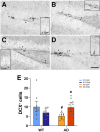
Fig 3.. Ctsb treatment improves adult neurogenesis in AD mice.
(A-D) Representative photomicrographs of the dentate gyrus derived from (A) WT-Con; (B) WT-Ctsb; (C) AD-Con; (D) AD-Ctsb brain tissue sections subjected to DCX staining. (E) Treatment with Ctsb in AD mice restored DCX cell number to WT-Con levels. (N; WT-Con = 10, WT-Ctsb = 8, AD-Con = 8, AD-Ctsb = 13). Scale bars: 50 μm overview; 25 μm inset. #P<0.05 AD compared to WT in the same Treatment. *P<0.05 Ctsb compared to Con in the same Genotype. Data in E was analyzed by GLM and are presented as Estimated Marginal Means and their respective 95% CI.
Fig 4.
Fig 4.. Ctsb treatment does not change amyloid plaque deposition.
(A,B) Representative photomicrographs of ThioS staining (green) in sections derived from (A) AD-Con and (B) AD-Ctsb brain tissue. Scale bar: 500 μm. (C-F) There was no difference between AD-Ctsb and AD-Con mice in ThioS+ amyloid plaque density or number in (C,D) cortex or (E,F) hippocampus. Data were analyzed by GLM and are presented as Estimated Marginal Means and their respective 95% CI. (N; AD-Con = 8, AD-Ctsb = 11), Cortex (CTX); Dentate gyrus (DG).
Fig 5.
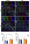
Fig 5.. Ctsb treatment does not modify neuroinflammation.
Representative photomicrographs of dorsal hippocampal sections subjected to GFAP (green), Iba1 (red) and DAPI (blue) labeling derived from (A) WT-Con, (B) WT-Ctsb, (C) AD-Con and (D) AD-Ctsb mice. (E,F) Astrocyte and microglia area density. Increased hippocampal density of (E) Iba1+ microglia but not of (F) GFAP+ astrocytes was observed in the AD groups. (N; WT-Con = 9, WT-Ctsb = 9, AD-Con = 8, AD-Ctsb = 12). Scale bars: 100 μm. *P<0.05 for Genotype effect (WT vs AD). Data were analyzed by GLM and are presented as Estimated Marginal Means and their respective 95% CI.
Fig 6.
Fig 6.. Ctsb treatment induces cytoskeletal reorganization in WT mice whereas transcription and RNA processing in AD mice.
(A) Volcano plot of average effect of AD. Gene set enrichment analysis (GSEA) based on (B) biological processes and (C) cellular components. (D) Scatter plots of proteins associated with the lysosome ontology that has log fold changes > 0.50. (E) Volcano plots showing significant proteins in pairwise group comparisons. (F) GSEA of biological processes summarizing changes observed in all groups. (G) Mean abundance of proteins associated with mRNA metabolic process ontology. (H) Volcano plot of interaction between AD and Ctsb treatment. (I) Mean abundance of proteins associated with myofibril and cytosolic ribosome ontologies, which were found to be enriched by GSEA of interaction data, using cellular components. (J) Scatter plots of selected proteins from our data that play roles in neurogenesis and neural plasticity. Multiple hypothesis testing was performed by Benjamini-Hochberg method for each differential expression analysis and proteins with FDR < 0.10 were considered statistically significant. For GSEAs, a cut-off of FDR < 0.05 was used.
Fig 7.
Fig 7.. Effects of Ctsb treatment on muscle and plasma proteome.
Muscle proteome: (A) GSEA plot of Wnt signaling pathway, as one of the most affected gene set in AD. GSEA of average effect of Ctsb treatment based on (B) biological processes. (C) GSEA using biological processes summarizing changes observed in each group. Plasma proteome: GSEA of (D) biological processes summarizing changes observed in the average effect of AD. (E) GSEA of biological processes summarizing changes observed in all groups. (F) Mean abundance of proteins associated with metabolic and cytoskeletal ontologies were displayed from GSEA of interaction data. (C,E,F) Data for the pairwise comparisons are annotated as follows: AD-WT(Ctsb), AD-Ctsb vs WT-Ctsb; AD-WT(Con), AD-Con vs WT-Con; Ctsb-Con(WT), WT-Ctsb vs WT-Con; Ctsb-Con(AD), AD-Ctsb vs AD-Con.

References

    1. Li X., et al., Global, regional, and national burden of Alzheimer’s disease and other dementias, 1990–2019. Front Aging Neurosci, 2022. 14: p. 937486. - PMC - PubMed
    1. Perneczky R., et al., Anti-amyloid antibody treatments for Alzheimer’s disease. Eur J Neurol, 2024. 31(2): p. e16049. - PMC - PubMed
    1. DeTure M.A. and Dickson D.W., The neuropathological diagnosis of Alzheimer’s disease. Mol Neurodegener, 2019. 14(1): p. 32. - PMC - PubMed
    1. Boyle P.A., et al., Association of muscle strength with the risk of Alzheimer disease and the rate of cognitive decline in community-dwelling older persons. Arch Neurol, 2009. 66(11): p. 1339–44. - PMC - PubMed
    1. Kerr N.R., et al., Hindlimb immobilization induces insulin resistance and elevates mitochondrial ROS production in the hippocampus of female rats. J Appl Physiol (1985), 2024. - PMC - PubMed

Publication types

LinkOut - more resources