[Preprint]. 2025 Jan 22:2025.01.17.633651.
doi: 10.1101/2025.01.17.633651.

Markovian State Models uncover Casein Kinase 1 dynamics that govern circadian period

Affiliations

Markovian State Models uncover Casein Kinase 1 dynamics that govern circadian period

Clarisse Gravina Ricci et al. bioRxiv. .

Abstract

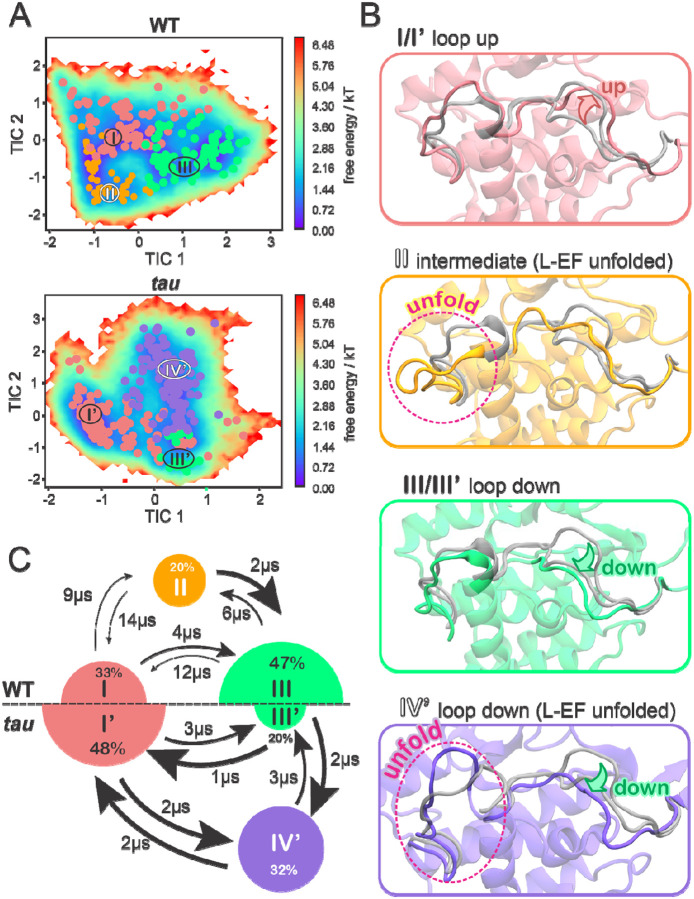
Circadian rhythms in mammals are tightly regulated through phosphorylation of Period (PER) proteins by Casein Kinase 1 (CK1, subtypes δ and ε). CK1 acts on at least two different regions of PER with opposing effects: phosphorylation of phosphodegron (pD) regions leads to PER degradation, while phosphorylation of the Familial Advanced Sleep Phase (FASP) region leads to PER stabilization. To investigate how substrate selectivity is encoded by the conformational dynamics of CK1, we performed a large set of independent molecular dynamics (MD) simulations of wildtype CK1 and the tau mutant (R178C) that biases kinase activity toward a pD. We used Markovian State Models (MSMs) to integrate the simulations into a single model of the conformational landscape of CK1 and used Gaussian accelerated molecular dynamics (GaMD) to build the first molecular model of CK1 and the unphosphorylated FASP motif. Together, these findings provide a mechanistic view of CK1, establishing how the activation loop acts as a key molecular switch to control substrate selectivity. We show that the tau mutant favors an alternative conformation of the activation loop and significantly accelerates the dynamics of CK1. This reshapes the binding cleft in a way that impairs FASP binding and would ultimately lead to PER destabilization and shorter circadian periods. Finally, we identified an allosteric pocket that could be targeted to bias this molecular switch. Our integrated approach offers a detailed model of CK1's conformational landscape and its relevance to normal, mutant, and druggable circadian timekeeping.


Conflict of interest statement

Declaration of interests The authors do not declare any conflicts of interest.

Figures

Figure 1.
Figure 1.
The CK1 activation loop acts as a molecular switch to control PER phosphorylation and circadian period. A) Simple schematic view of the transcriptional-translational feedback loop at the core of the human clock. B) The circadian period is finely regulated by a phosphoswitch mechanism controlling PER stability. C) Apo structure of CK1δ (PDB 1CKJ), highlighting: the location of the catalytic HRD motif (red star); the two conserved anion binding sites (purple circles), and the alternative conformations of the activation loop (pink and green). D) Sulfate anions bind to the first anion site via two positively charged clamps (R178 and K224). The R178C mutation in tau impairs the ability of this site to bind anions. E) Alternative conformations of the activation loop in tau (PDB 6PXN), showing that the loop up conformation (pink) sterically blocks the second anion site.
Figure 2.
Figure 2.
The equilibrium between three preferred conformational states is accelerated and inverted by the CK1 tau mutant. A) Free energy landscapes of the WT CK1 (top) and tau mutant (bottom) in terms of the slowest tICA components, with microstates clustered into meta-stable states (identified by roman numerals). B) Representative conformations of each meta-stable state. For comparison, x-ray conformations of the activation loop and L-EF are superimposed (transparent gray; loop down PDB 1CKJ, loop up PDB 6PXN). C) Equilibrium populations of meta-stable states and MFPTs between states for the WT (top) and tau (bottom) protein systems. Radius of the circles is proportional to the equilibrium population percentage and thickness of the arrows is proportional to transition rates between states. The numbers next to the arrows indicate MFPTs.
Figure 3.
Figure 3.
The role of Gly175 for the conformational dynamics of the activation loop. A) View of x-ray structures highlighting the different configurations adopted by Gly175 in ‘loop up’ and ‘loop down’ conformations of the CK1 activation loop (PDB 6PXN and 1CKJ, respectively). B) Schematic representation of the conformational landscape involving the activation loop, based on MSM-derived kinetics and stability of the two most populated states in each system (WT CK1, top; and tau, bottom).
Figure 4.
Figure 4.
Molecular model of the interaction between CK1 and unphosphorylated FASP. A) The FASP region of human PER2. Residue numbers with arched arrows represent serines that are sequentially phosphorylated by CK1. Positions listed below (−4 – +5) are relative to priming serine, S662. B) Representative structure of the final model with the unphosphorylated FASP peptide (purple) and CK1 (green). For clarity, we use one-letter amino acid code for residues of the substrate (FASP) and three-letter code for residues of the kinase CK1. Pink star represents the priming event of S662. C) Atomic fluctuations of the bound FASP based on accumulated molecular dynamics trajectories. D-F) Distance-based interaction histograms involving V663 at position +1 (panel D), E661 at position −1 (panel E), and K659 at position −3 (panel F) with CK1 residues or ATP.
Figure 5.
Figure 5.
Biochemical validation of the model for non-consensus FASP priming. A) NMR-based timecourse kinase assay quantifying the increase in peak volume corresponding to the phosphopeak of the priming serine in human PER2 FASP (S662), taken from a series of 15N-1H HSQC spectra (Figure S14). B) ADP-Glo titration assay comparing WT FASP and alanine mutants at positions −3, −1, and +1 in the ‘priming only’ background (S665A mutation to halt sequential kinase activity, see Figure S15A). C) Quantification of the mean and SD of the KM from replicate titration experiments as shown in panel B, n=2. D) 15N-1H HSQC spectra comparing a ‘priming disrupted’ FASP peptide (S662A, black) and a ‘priming rescued’ FASP peptide (S662A/L666V, teal) in the presence of CK1. Zoom shows the region of the spectra where phosphoserines appear. E) Zoom of co-crystal structure of CK1 and a triply phosphorylated Tap63α PAD peptide, 3pPAD, highlighting the active site and +1 hydrophobic pocket region (PDB 6RU8). The +1 valine residue is inserted between the small hydrophobic pocket created between Tyr225 and Leu173 of CK1 when the activation loop is in the downward conformation. F) ADP-Glo titration of the ‘priming only’ (S665A) FASP peptide comparing CK1 WT (black) and D132A (purple).
Figure 6.
Figure 6.
The conformation of the activation loop shapes the substrate binding cleft. A) The ‘loop down’ conformation of the activation loop creates a straight binding cleft whereas (B) the ‘loop up’ conformation creates a bent binding cleft, opening a sub-pocket right under the active site. The solid blobs represent density maps computed based on organic probes from FTMap while the meshed blobs represent occupancy maps computed for residues Lys171, Asn172 and Leu173 along the simulations. C-D) Co-crystal structure of human PER2 pFASP bound to CK1 (PDB 8D7M) in the straight substrate binding cleft (C), whereas the dPER perShort peptide binds to CK1 (PDB 8D7P) following a bent substrate binding cleft (D).
Figure 7.
Figure 7.
A potential allosteric pocket to shorten the circadian rhythm. A) Residues belonging to the tau activation pocket are highlighted in yellow, while the small organic molecules used as probes by FTMap are represented as grey sticks. B) Complete assembly of this pocket occurs when the activation loop is ‘up’. Ligand binding to this pocket is likely to stabilize the ‘loop up’ conformation.

References

    1. Aschoff J., Circadian Rhythms in Man. Science, 1965. 148(3676): p. 1427–32. - PubMed
    1. Rosbash M., Molecular Control of Circadian-Rhythms. Current Opinion in Genetics & Development, 1995. 5(5): p. 662–668. - PubMed
    1. Partch C.L., Green C.B., and Takahashi J.S., Molecular architecture of the mammalian circadian clock. Trends Cell Biol, 2014. 24(2): p. 90–9. - PMC - PubMed
    1. Cohen S.E. and Golden S.S., Circadian Rhythms in Cyanobacteria. Microbiol Mol Biol Rev, 2015. 79(4): p. 373–85. - PMC - PubMed
    1. Eelderink-Chen Z., et al., A circadian clock in a nonphotosynthetic prokaryote. Sci Adv, 2021. 7(2). - PMC - PubMed

Publication types