[Preprint]. 2025 Jan 22:2025.01.20.633914.
doi: 10.1101/2025.01.20.633914.

SLAMF6 enables efficient attachment, synapse formation, and killing of HIV-1-infected CD4+ T cells by virus-specific CD8+ T cells

Affiliations

SLAMF6 enables efficient attachment, synapse formation, and killing of HIV-1-infected CD4+ T cells by virus-specific CD8+ T cells

Blandine Monel et al. bioRxiv. .

Abstract

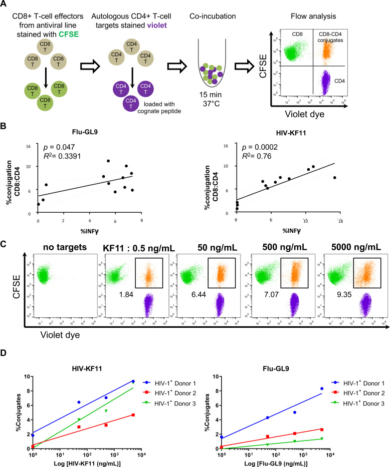
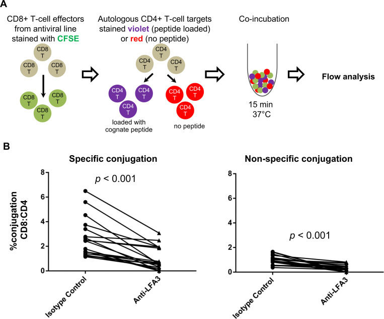
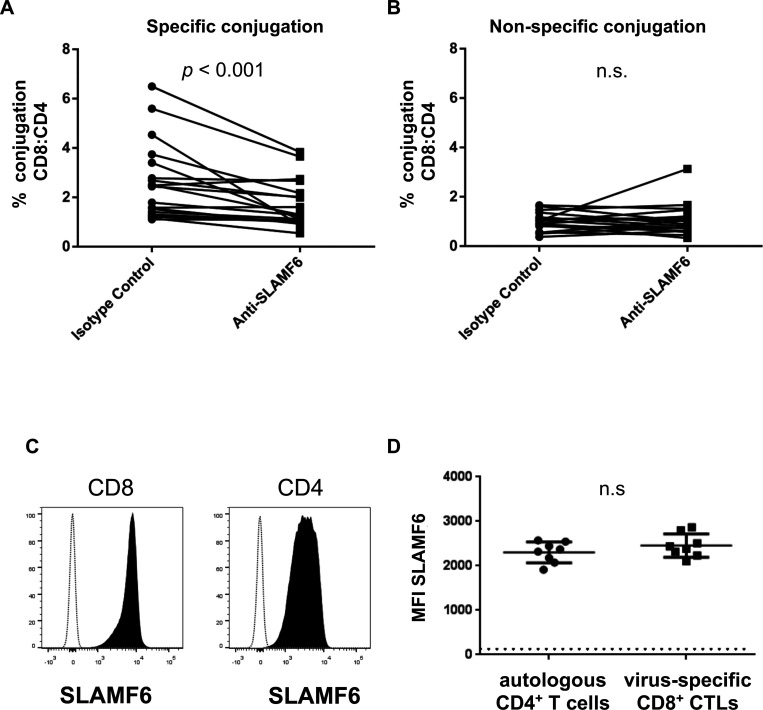
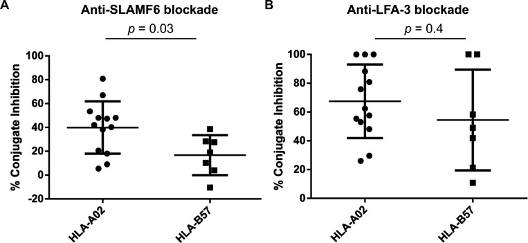
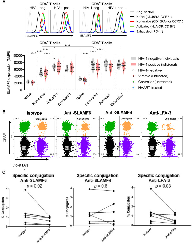
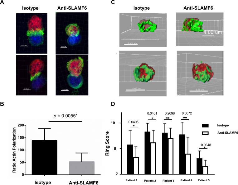
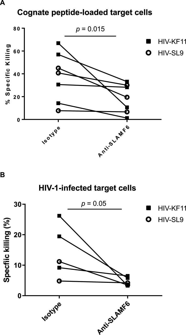
Efficient recognition and elimination of HIV-1-infected CD4+ T cells by cytotoxic CD8+ T cells (CTLs) require target cell engagement and the formation of a well-organized immunological synapse. Surface proteins belonging to the SLAM family are known to be crucial for stabilizing the immunological synapse and regulating antiviral responses during lymphotropic viral infections. In the context of HIV-1, there have been reports of SLAMF6 down-regulation in HIV-1-infected CD4+ T cells; however, the significance of this modulation for CTL function remains unclear. In this investigation, we used CTL lines from People living with HIV (PLWH) to examine the impact of SLAMF6 blockade on three pivotal processes: (1) the formation of CD8+-CD4+ T-cell conjugates, (2) the establishment of the immunological synapse, and (3) the killing and cytokine production capacity of HIV-1-specific CTLs during HIV-1 infection. Our findings reveal that the inability to form CD8+-CD4+ T-cell conjugates following incubation with an anti-SLAMF6 blocking antibody is primarily attributable to a defect in actin ring formation at the immunological synapse. Furthermore, SLAMF6 blockade leads to a reduction in the killing efficiency of HIV-1-infected CD4+ T cells by HIV-1-specific CTLs, underscoring the critical role of SLAMF6 in cytolytic function. This study highlights the importance of SLAMF6 receptors in modulating cytotoxic antiviral responses, shedding light on potential avenues for manipulation and enhancement of this pathway in the context of HIV and other lymphotropic viral infections.

Keywords: Cytokine production; Cytotoxic T lymphocytes (CTLs); Cytotoxic immune response; HIV-1; Immunological synapse; SLAMF6.


Figures

Figure 1:
Figure 1:. Conjugation of virus-specific CD8+ CTLs to CD4+ T-cell targets loaded with cognate peptide can be accurately measured via flow cytometry and predict CTL functionality.
(A) Experimental two-color design for quantifying antigen-specific CD8+ and CD4+ T-cell Interactions is shown. In this experiment, CTLs were labeled with green fluorescence (CFSE) and autologous CD4+ T cells were loaded with varying doses of cognate peptide and stained with a violet dye. Effector and target cells were mixed at a 1:1 ratio of CD8+ to CD4+ T cells and co-cultured at 37°C for 15 minutes. Subsequently, the cells were fixed and subjected to flow cytometry analysis. The rate of antigen-specific conjugations was determined by calculating the frequency of double-positive cells, where green effector cells overlapped with violet target cells, located in the upper right quadrant of the cytometry plot. (B) We investigated the relationship between the frequency of CD8+-CD4+ T cell conjugate formation occurring within a 15-minute timeframe and subsequent cytokine production by CD8+ CTLs at six hours of co-culture. This was done for HIV-1 KF11 peptide (HIV-KF11)-specific CTLs (left) and influenza GL9 peptide (Flu-GL9)-specific CTLs (right) at varying concentration of cognate peptide loaded onto autologous CD4+ T-cell targets (n = 12 for each). The X-axis represents the frequency of specific conjugate formation observed after 15 minutes, while the Y-axis illustrates the frequency of IFN-γ+ CTLs after 6 hours of co-culture. The graphs presented here serve as representatives from a total of three independent experiments, and the correlation between these variables was assessed using a simple linear regression (p values and R2 shown on graphs). (C) CD4+ T cell targets were loaded with the HIV-KF11 epitope at the indicated concentrations and then washed to remove excess peptide. HIV-KF11-specific CD8+ CTLs were incubated with autologous peptide-pulsed CD4+ T cell targets for 15 minutes. Each flow plot represents a different concentration of the loaded peptide, and the conjugate formation is depicted in orange (identified as the double-positive green and violet population) and the conjugate frequency is indicated for each plot. This experiment is representative of three independent experiments. (D) The effect of cognate peptide concentration on conjugates formation was measured by co-incubating HIV-KF11-specific (left) or Flu-GL9-specific (right) CTLs with autologous CD4+ T-cell targets loaded with different concentration of HIV-KF11 or Flu-GL9 peptide. A simple linear regression between the log-transformed peptide concentration (X-axis) and percent of conjugates (Y-axis) was assessed, with results as follows: KF11 Donor 1 (R2 = 0.96, p = 0.02), KF11 Donor 2 (R2 = 0.95, p = 0.02), KF11 Donor 3 (R2 = 0.96, p = 0.02), GL9 Donor 1 (R2 = 0.94, p = 0.03), GL9 Donor 2 (R2 = 0.97, p = 0.01), and GL9 Donor 3 (R2 = 0.99, p = 0.006).
Figure 2:
Figure 2:. Blockade of LFA-3 inhibits both antigen-specific and non-specific attachment and conjugation of CD8+ CTLs to autologous CD4+ T cells.
(A) We developed a three-colors experimental design for measuring antigen specific and non-specific attachment and conjugation of CD8+ CTLs to autologous CD4+ T cells. CTLs were labeled with green fluorescence (CFSE) and autologous CD4+ T cells isolated from donor PBMCs were divided into two samples: peptide-pulsed targets cells stained in violet and non-peptide-pulsed target cells stained in red. Peptide-pulsed target cells and non-pulsed target cells were co-incubated at a ratio 1:1:1 for 15 minutes at 37°C. Cells were fixed and analyzed by flow cytometry. Within the CFSE+ cells (i.e., CD8+ CTLs) the rate of antigen-specific conjugation is determined by the frequency of violet-positive conjugates and the rate of non-specific conjugation is determined by the frequency of red-positive conjugates. (B) CD8+-CD4+ conjugate formation sensitive to LFA-3 blockade was explored. CD8+ T cells were co-cultured with CD4+ T target cells for 15 minutes. Conjugate formation was analyzed by flow cytometry, gating on cells that were double positive for CD8 and CD4. Two conditions were assessed: (i) Percent of conjugation of CD8+ T cells (CFSE+) and CD4+ T cells loaded with cognate peptide (Violet+) (left panel), and (ii) Percent of conjugation of CD8+ T cells (CFSE+) and CD4+ T cells not loaded with cognate peptide (Red+) (right panel). The analysis was performed using either an isotype control antibody or a blocking antibody against LFA-3 (CD58). Co-cultures were analyzed to determine the percentage of specific and non-specific conjugates formed under these conditions. Data represents the conjugation rate from 20 independent experiments (n = 20).
Figure 3:
Figure 3:. SLAMF6 is required for efficient formation of primary human antiviral CD8+-CD4+ T cell conjugates.
(A-B) The conjugation assay was performed with primary T cell lines from a cohort of HIV1-positive subjects (n = 10). Multiple CTL lines were generated for each donor and were specific for any of the following viral epitopes: HIV-SL9; HIV-KF11; Flu-GL9. Compiled results from a total of 20 CTL lines are shown. The rate of specific conjugation in the presence of SLAMF6 blockade or isotype control is shown in (A) and the rate of non-specific conjugation is shown in (B) The effect of the blockade was compared to the IgG isotype control by a two-tailed Wilcoxon matched-pairs signed rank test; p values are shown on graph, with n.s. denoting non-significant (p > 0.05). (C) Representative flow cytometry histograms measuring SLAMF6 expression on T cells used for the conjugation assay is shown for virus-specific CD8+ CTLs (left) and autologous primary CD4+ T cells isolated from PBMC and maintained for 3 days in medium containing IL-2 (50 U/ml) (right). The black-filled histogram represents SLAMF6 staining and the dotted histogram represents the FMO (fluorescence minus one). (D) Combined results of SLAMF6 expression on CTLs and IL-2-cultured CD4+ T cells from 10 different HIV-1-positive subjects are shown. Two-tailed Wilcoxon matched-pairs signed rank test showed no statistically significant differences (n.s.).
Figure 4:
Figure 4:. HLA-B57:01-restricted CTL lines are less sensitive to SLAMF6 blockade than HLA-A*02:01-restricted lines.
For each CTL line studied, the “percent conjugate inhibition” was calculated as: 100% - (Percent specific conjugation in the presence of the blockade / Percent of specific conjugation in the presence of the IgG Isotype control). The mean “specific-conjugate inhibition” in the presence of blockade of (A) SLAMF6 or (B) LFA-3 was assessed for HLA-B57:01-restricted CTL lines (n = 7) compared to HLA-A*02:01-restricted CTL lines (n = 13) and compared by the Mann-Whitney test. The p-value is indicated for each graph.
Figure 5:
Figure 5:. SLAMF6 is highly but differentially expressed on CD8+ and CD4+ T-cell subsets and is required for efficient CD8+-CD4+ T cell conjugate formation directly ex vivo.
(A) SLAMF6 expression, measured as median fluorescence intensity (MdFI), was assessed on different CD8+ T and CD4+ T-cell subsets in PBMCs from HIV-1-positive (n = 10) and HIV-1-negative (n = 10) donors ex vivo via flow cytometry; representative histograms of SLAMF6 expression on different T-cell subsets is shown, with aggregate data plotted below as violin plots. One-way ANOVA with correction for multiple comparisons was performed; statistical significance is denoted as follows: * p < 0.05, ** p < 0.01, **** p < 0.0001. (B-C). Conjugation assay on CD8+ T cells from ex vivo PBMCs and autologous CD4+ T target cells in the presence of SLAMF6, SLAMF4 or LFA-3 blocking antibodies, or isotype IgG control. The peptides used in this assay were pools of overlapping synthetic peptides representing the HIV-1 Gag protein or the CMV pp65 protein. Specific conjugation is shown in the upper left quadrant. All subjects studied were HIV-1 and CMV co-infected. The flow plots in B are representative of six additional conjugation assays performed on ex vivo T cells. The rate of specific conjugation in the presence of a SLAMF6, SLAMF4 and LFA-3 blockade was assessed and shown in C (n = 6). The effects of the blockade were compared to an IgG isotype control using a two-tailed Wilcoxon matched-pairs signed rank test with p-values shown on graphs.
Figure 6:
Figure 6:. SLAMF6 is necessary for immunological synapse formation between CD8+ T effector cells and CD4+ T target cells.
(A) HIV-KF11-specific CD8+ T cells (red) were co-cultured for fifteen minutes with autologous antigen-loaded CD4+ T cell targets (blue) in the absence or presence of a SLAMF6 blockade, stained for actin localization with phalloidin (green), and analyzed by confocal microscopy. Four different images with a magnification of X100 are shown, representative of 100 different pictures taken. (B) Antigen-specific actin polarization in CD8+-CD4+ T cell contacts is observed in the presence of the SLAMF6 blockade (right) and isotype control (left). The ratio of actin polarization in the presence or absence of the SLAMF6 blockade was compared using the Mann-Whitney test; the p-value is indicated in the graph. (C) The 3D plots show en face views of the CD4+-CD8+ T cell contacts with a magnification of 100X. The blue pixels were removed to allow an unobstructed view of the synapse. Four representative images are shown, both in the presence of the isotype control (left) and the SLAMF6 blockade (right). (D) The ring formation was scored for all 100 images analyzed from 5 independent experiments, and SLAMF6 vs. isotype control were compared for each experiment and analyzed by the Mann-Whitney test; the p-value is indicated in the graph.
Figure 7:
Figure 7:. SLAMF6 is necessary for CD8+ T cell killing function.
(A) Cr 51-labeled CD4+ T cells were loaded with two different peptides, either HIV-KF11 (black squares) or HIV-SL9 (open circles), at 5 μg/mL and co-cultured with autologous peptide-specific CD8+ T cells. Cr 51 release from effectors was assessed in the presence of a SLAMF6 blocking antibody or an isotype control. Seven different CTL lines were tested. (B) Activated primary CD4+ T cells were infected with HIV-1 (clone NL4–3) virus for 48 hours, then labeled with Cr 51 and incubated with autologous peptide-specific CD8+ T effector cells for 4 hours at a ratio of 1:1 in the presence of a SLAMF6 blockade or an isotype control. Five different CTL lines were tested. Black squares represent the HIV-KF11-specific CTL lines, and open circles represent the HIV-SL9-specific CTL lines tested. Specific killing of target cells in the Cr 51 release assay was calculated as 100x (sample release – spontaneous release) / (maximum release - spontaneous release). Statistical differences were measured using the Wilcoxon matched-pairs test with p-values shown on graph.

Similar articles

References

    1. Streeck H., Jolin J. S., Qi Y., Yassine-Diab B., Johnson R. C., Kwon D. S., Addo M. M., Brumme C., Routy J.-P., Little S., Jessen H. K., Kelleher A. D., Hecht F. M., Sekaly R.-P., Rosenberg E. S., Walker B. D., Carrington M., and Altfeld M.. 2009. Human immunodeficiency virus type 1-specific CD8+ T-cell responses during primary infection are major determinants of the viral set point and loss of CD4+ T cells. J Virol 83: 7641–8. - PMC - PubMed
    1. Schmitz J. E., Kuroda M. J., Santra S., Sasseville V. G., Simon M. A., Lifton M. A., Racz P., Tenner-Racz K., Dalesandro M., Scallon B. J., Ghrayeb J., Forman M. A., Montefiori D. C., Rieber E. P., Letvin N. L., and Reimann K. A.. 1999. Control of viremia in simian immunodeficiency virus infection by CD8+ lymphocytes. Science 283: 857–60. - PubMed
    1. Schmitz J. E. 1999. Control of Viremia in Simian Immunodeficiency Virus Infection by CD8+ Lymphocytes. Science (1979) 283: 857–860. - PubMed
    1. Lim S. Y., Lee J., Osuna C. E., Vikhe P., Schalk D. R., Chen E., Fray E., Kumar M., Schultz-Darken N., Rakasz E., Capuano S., Ladd R. A., Gil H. M., Evans D. T., Jeng E. K., Seaman M., Martin M., Van Dorp C., Perelson A. S., Wong H. C., Siliciano J. D., Siliciano R., Safrit J. T., Nixon D. F., Soon-Shiong P., Nussenzweig M., and Whitney J. B.. 2024. Induction of durable remission by dual immunotherapy in SHIV-infected ART-suppressed macaques. Science 383: 1104–1111. - PMC - PubMed
    1. Seller Z. 2001. Cellular Adhesion and Adhesion Molecules. Turkish Journal of Biology 25: 1–15.

Publication types