[Preprint]. 2025 Jan 24:2024.12.18.629302.
doi: 10.1101/2024.12.18.629302.

Mettl15-Mettl17 modulates the transition from early to late pre-mitoribosome

Affiliations

Mettl15-Mettl17 modulates the transition from early to late pre-mitoribosome

Yury Zgadzay et al. bioRxiv. .

Abstract

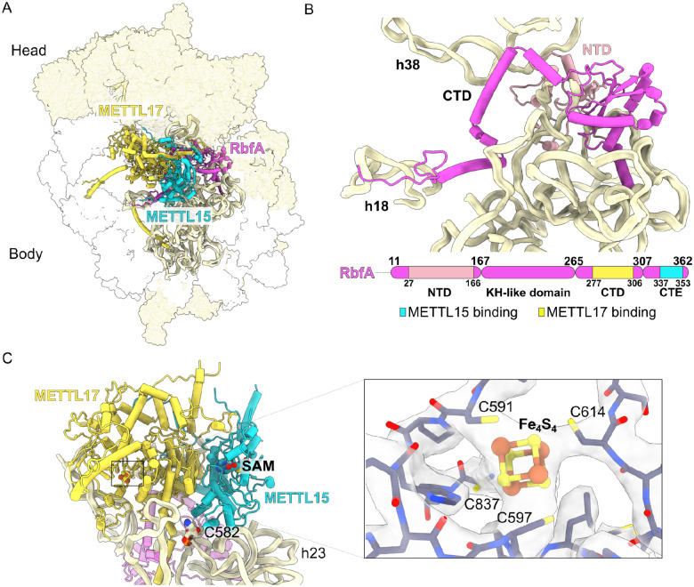
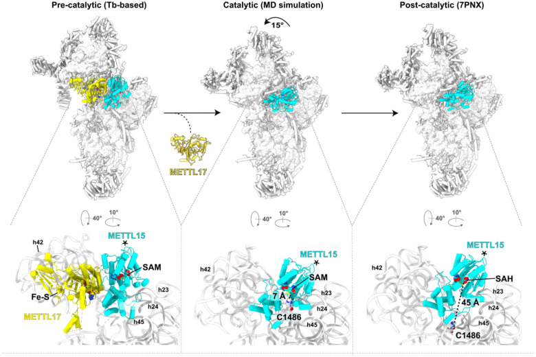
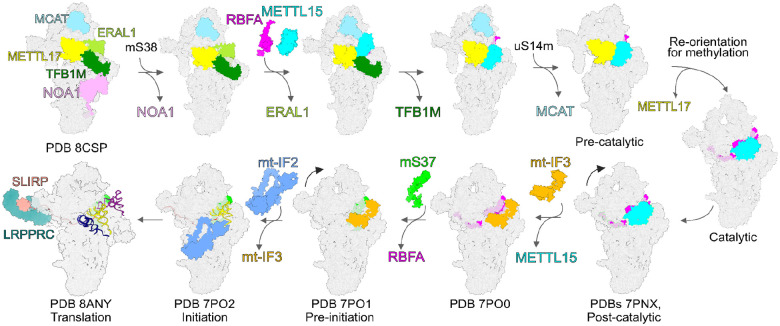
The assembly of the mitoribosomal small subunit involves folding and modification of rRNA, and its association with mitoribosomal proteins. This process is assisted by a dynamic network of assembly factors. Conserved methyltransferases Mettl15 and Mettl17 act on the solvent-exposed surface of rRNA. Binding of Mettl17 is associated with the early assembly stage, whereas Mettl15 is involved in the late stage, but the mechanism of transition between the two was unclear. Here, we integrate structural data from Trypanosoma brucei with mammalian homologs and molecular dynamics simulations. We reveal how the interplay of Mettl15 and Mettl17 in intermediate steps links the distinct stages of small subunit assembly. The analysis suggests a model wherein Mettl17 acts as a platform for Mettl15 recruitment. Subsequent release of Mettl17 allows a conformational change of Mettl15 for substrate recognition. Upon methylation, Mettl15 adopts a loosely bound state which ultimately leads to its replacement by initiation factors, concluding the assembly. Together, our results indicate that assembly factors Mettl15 and Mettl17 cooperate to regulate the biogenesis process, and present a structural data resource for understanding molecular adaptations of assembly factors in mitoribosome.


Figures

Figure 1.
Figure 1.. Dimer of Mettl15 and Mettl17 is found with RbfA in a T. brucei pre-mtSSU.
(A) Structure of pre-mtSSU highlights Mettl15 (yellow), Mettl17 (cyan), RbfA (magenta) and rRNA (pale-yellow ribbon). Other assembly factors and mitoribosomal proteins are shown as white and yellow surfaces, respectively. (B) Structure and schematic representation of RbfA and its interaction with rRNA. Mettl15 and Mettl17 interacting regions are indicated. (C) Structure of the Mettl15-Mettl17 heterodimer with cofactors. A close-up view shows Fe4S4 with its coordinating residues.
Figure 2.
Figure 2.. Mettl17 and Mettl15 are widely distributed across eukaryotes.
Distribution in major eukaryotic groups mapped onto a phylogenetic tree. The numbers in parentheses indicate the number of organisms searched in the respective group. Filled symbols represent presence in all organisms, half-filled symbols indicate presence in a subset of organisms, and empty symbols indicate absence.
Figure 3:
Figure 3:. Modelling of early pre-mtSSU with Mettl15.
(A) AlphaFold model of Mettl17-TFB1M superposed with the experimentally determined model of the early state (grey, PDB 8CSP). (B) AlphaFold model of Mettl15-TFB1M. (C) Left, model of early pre-mtSSU with all three methyltransferases, including Mettl15. Right, schematics of protein-protein interactions of methyltransferases, other assembly factors (colored nodes), and mitoribosomal proteins (grey nodes). The node size corresponds to relative molecular mass of protein subunits, and the connector width corresponds to the relative solvent accessible interface area buried between the subunits, calculated with PDBePISA v.1.52.
Figure 4:
Figure 4:. Simulation reveals low-energy structural fluctuations about post-catalytic configuration.
(A) Comparison of Mettl15 orientations between catalytic (cyan) and post-catalytic (smoky blue) states based on superposition of the mtSSU. Shifts between equivalent Mettl15 Cα atoms and rRNA phosphorus atoms in the different states are color-coded using the spectrum from dark blue to red, corresponding to the range from 0 to 20 Å. (B) A.s.d of Mettl15 calculated with respect to the post-catalytic conformation. There are no major intramolecular structural deformations required to adopt orientations in which SAM is proximal to C1486 (labeled “restrained”). The most notable difference is an increase of ~0.5 Å in Leu274. (C) Comparison of Mettl15 orientations between pre-catalytic (brown) and catalytic (cyan) states. The angles describing Mettl15 rotation between the states are indicated. (D) Free energy of Mettl15, calculated from a simulation without SAM-C1486 restraints (i.e. unrestrained). When the restraint is included, short distances between SAM and C1486 (<7 Å) can be reached when Mettl15 is rotated/tilted by ~7–12°. Each “x” indicates a simulated conformation in which the distance is small (<7 Å) in the restrained simulations. These domain orientations are associated with small increases in free energy, indicating that thermal energy is sufficient for Mettl15 to spontaneously adopt catalytically-compatible poses.
Figure 5:
Figure 5:. Sequential steps assembly with Mettl17 and Mettl15.
Left, T. brucei based model of human pre-mtSSU (PDB ID 8CST) with Mettl17-Mettl15. Middle, model from molecular dynamics simulations with rearranged Mettl15 bringing it to the substrate for its methylation. Right, model of the post-catalytic state (PDB ID 7PNX), in which the target nucleotide C1486 is resolved. The estimated distances between the target nucleotide and Mettl15 cofactor SAM are shown and selected rRNA helices are annotated in the close-up views. Asterisks indicate equivalent elements in the three states.
Figure 6:
Figure 6:. Proposed mtSSU pathway with precursors containing Mettl15-Mettl17 heterodimer and the pre-mtSSU with three methyltransferases.
Assembly and initiation factors are shown as colored-coded surfaces. Mitoribosomal proteins and RNA are shown as grey surfaces.

Similar articles

References

    1. Singh V. et al. Structural basis of LRPPRC-SLIRP-dependent translation by the mitoribosome. Nat Struct Mol Biol (2024). - PMC - PubMed
    1. Itoh Y. et al. Mechanism of membrane-tethered mitochondrial protein synthesis. Science 371, 846–849 (2021). - PMC - PubMed
    1. Ott M., Amunts A. & Brown A. Organization and Regulation of Mitochondrial Protein Synthesis. Annu Rev Biochem 85, 77–101 (2016). - PubMed
    1. Singh V. et al. Mitoribosome structure with cofactors and modifications reveals mechanism of ligand binding and interactions with L1 stalk. Nat Commun 15, 4272 (2024). - PMC - PubMed
    1. Amunts A., Brown A., Toots J., Scheres S.H.W. & Ramakrishnan V. Ribosome. The structure of the human mitochondrial ribosome. Science 348, 95–98 (2015). - PMC - PubMed

Publication types

LinkOut - more resources