[Preprint]. 2025 Apr 25:2025.01.17.633682.
doi: 10.1101/2025.01.17.633682.

Enantioselective Protein Affinity Selection Mass Spectrometry (E-ASMS)

Affiliations

Enantioselective Protein Affinity Selection Mass Spectrometry (E-ASMS)

Xiaoyun Wang et al. bioRxiv. .

Abstract

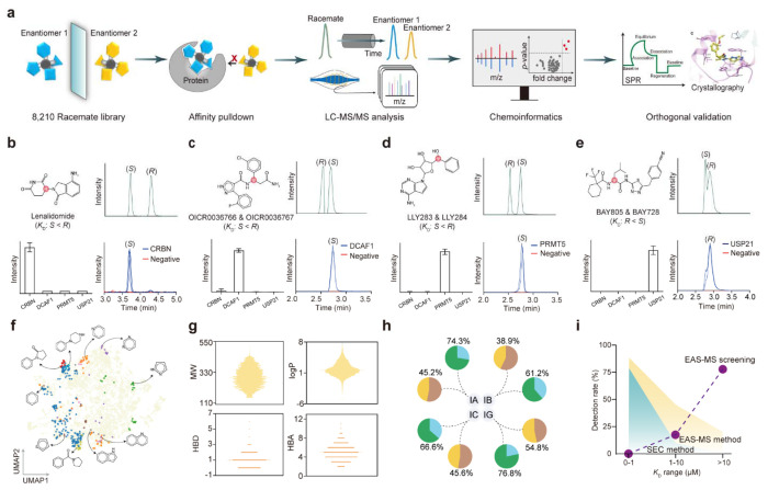
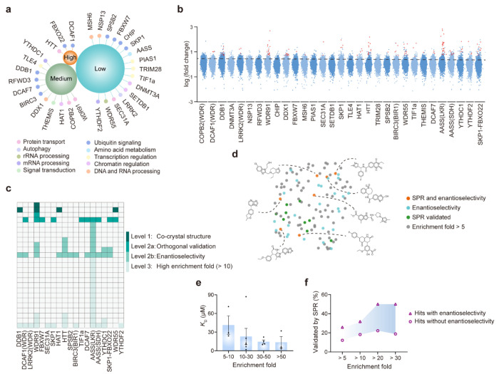
We report an enantioselective protein affinity selection mass spectrometry screening approach (E-ASMS) that enables the detection of weak binders, informs on selectivity, and generates orthogonal confirmation of binding. After method development with control proteins, we screened 31 human proteins against a designed library of 8,210 chiral compounds. 16 binders to 12 targets, including many proteins predicted to be "challenging to ligand", were discovered and confirmed in orthogonal biophysical assays. 7 binders to 6 targets bound in an enantioselective manner, with K D values ranging from 3 to 20 μM. Binders for four targets (DDB1, WDR91, WDR55, and HAT1) were selected for in-depth characterization using X-ray crystallography. In all four cases, the mechanism for enantioselectivity was readily explained. We conclude E-ASMS can be used to identify and characterize selective and weakly-binding ligands for novel protein targets with unprecedented throughput and sensitivity.


Conflict of interest statement

Competing interests The authors declare no competing interests.

Figures

Fig. 1 |
Fig. 1 |. Development of the E-ASMS platform for scalable protein ligandability discovery.
(a) Schematic representation of the E-ASMS screening workflow. (b-e) Benchmarking the E-ASMS platform using four positive control ligands binding to CRBN, DCAF1, PRMT5, and USP21, respectively. (f) Chemical space of the E-ASMS library. A two-dimensional uniform manifold approximation and projection (UMAP) visualization of SMILES descriptor. The displayed structures represent the top 10 scaffolds within the E-ASMS chemical library. (g) Molecular properties of the E-ASMS chemical library: molecular weight (MW), logP, hydrogen bond acceptors (HBA) and donors (HBD). (h) Chiral separation of the E-ASMS library using four chiral columns (IA, IB, IC, and IG), and two mobile phases (acetonitrile/methanol, yellow; water/methanol, green). (i) Detection rate of 109 ligands against 8 proteins by SEC and E-ASMS methods, respectively. Purple dots represent the KD distribution of chemical ligands discovered in the present E-ASMS screening against 31 proteins.
Fig. 2 |
Fig. 2 |. Scalable chemical ligand profiling across 31 diverse proteins.
(a) Overview of the 31 screened proteins. ‘High’: drug-like density (DLID) > 1, ‘Medium’: 0.5 < DLID < 1, ‘Low’: DLID < 0.5. (b) Scatter plot of all E-ASMS library compounds interacting with 31 proteins. Dots above the line represent hits with enrichment fold > 5. Red dots indicate hits with enrichment fold > 5 and p-value < 0.05. (c) Confidence levels of all identified hits according to orthogonal evidence. (d) UMAP visualization of the chemical space for hits with enrichment fold > 5. (e) Correlation between KD and enrichment fold. (f) SPR validation rate of hits with or without enantioselectivity, across different enrichment fold ranges.
Fig. 3 |
Fig. 3 |. Identification of enantioselective hits for DDB1, WDR91, WDR55, and HAT1.
(a) Scatter plot showing XS381952 pulled down by DDB1. (b) Chiral chromatograms of the XS381952 enantiomers before (upper) and after E-ASMS (bottom). (c) Co-crystal structure of DDB1 with XS381952. Hydrogen bond between the protein and the ligand is shown as green dashed line. (d) Scatter plot showing XS838489 pulled down by WDR91. (e) Chiral chromatograms of the XS838489 enantiomers. (f) Co-crystal structure of WDR91 with XS838489. Hydrogen bonds between the protein and the ligand are shown as green dashed lines. (g) Scat er plot showing XS381774 pulled down by WDR55. (h) Chiral chromatograms of the XS381774 enantiomers. (i) Co-crystal structure of WDR55 with XS381774. (j) Scatter plot showing XS380871 pulled down by HAT1. (k) Chiral chromatograms of the XS380871 enantiomers. (l) Co-crystal structure of HAT1 with XS380871. Hydrogen bonds between the protein and the ligand are shown as green dashed lines.

References

    1. Moffat J. G., Rudolph J. & Bailey D. Phenotypic screening in cancer drug discovery - past, present and future. Nature reviews. Drug discovery 13, 588–602 (2014). 10.1038/nrd4366 - DOI - PubMed
    1. Eder J., Sedrani R. & Wiesmann C. The discovery of first-in-class drugs: origins and evolution. Nature reviews. Drug discovery 13, 577–587 (2014). 10.1038/nrd4336 - DOI - PubMed
    1. Macarron R. et al. Impact of high-throughput screening in biomedical research. Nature reviews. Drug discovery 10, 188–195 (2011). 10.1038/nrd3368 - DOI - PubMed
    1. Parker C. G. et al. Ligand and target discovery by fragment-based screening in human cells. Cell 168, 527–541 e529 (2017). 10.1016/j.cell.2016.12.029 - DOI - PMC - PubMed
    1. Abbasov M. E. et al. A proteome-wide atlas of lysine-reactive chemistry. Nature chemistry 13, 1081–1092 (2021). 10.1038/s41557-021-00765-4 - DOI - PMC - PubMed

Publication types

LinkOut - more resources