. 2025 Jan 16:44:100945.
doi: 10.1016/j.bbih.2025.100945. eCollection 2025 Mar.

Glial activation among individuals with neurological post-acute sequelae of coronavirus disease 2019: A positron emission tomography study of brain fog using [18F]-FEPPA

Affiliations

Glial activation among individuals with neurological post-acute sequelae of coronavirus disease 2019: A positron emission tomography study of brain fog using [18F]-FEPPA

Sean A P Clouston et al. Brain Behav Immun Health. .

Abstract

Background: This study examined the regional distribution of glial activation in essential workers with neurological post-acute sequelae of coronavirus disease 2019 (COVID-19) infections (N-PASC).

Methods: We injected ≤185 MBq of [18F]-FEPPA as an intravenous bolus and positron-emission tomography over 2 h. To measure distribution volume (VT) we recruited 24 essential workers (14 N-PASC, 10 Never-COVID-19 Controls, of whom 22 successfully placed arterial lines). Individuals with low binding affinity were excluded from this study, and VT was adjusted for translocator protein genotype. Analyses that passed the false discovery rate are reported.

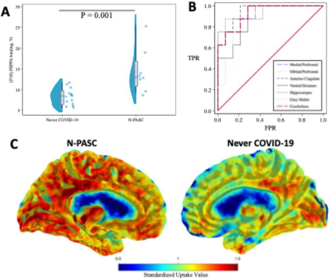
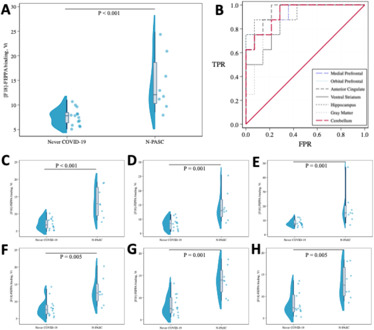
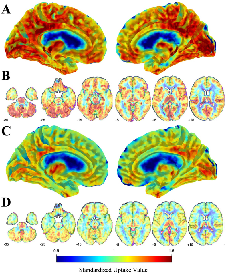
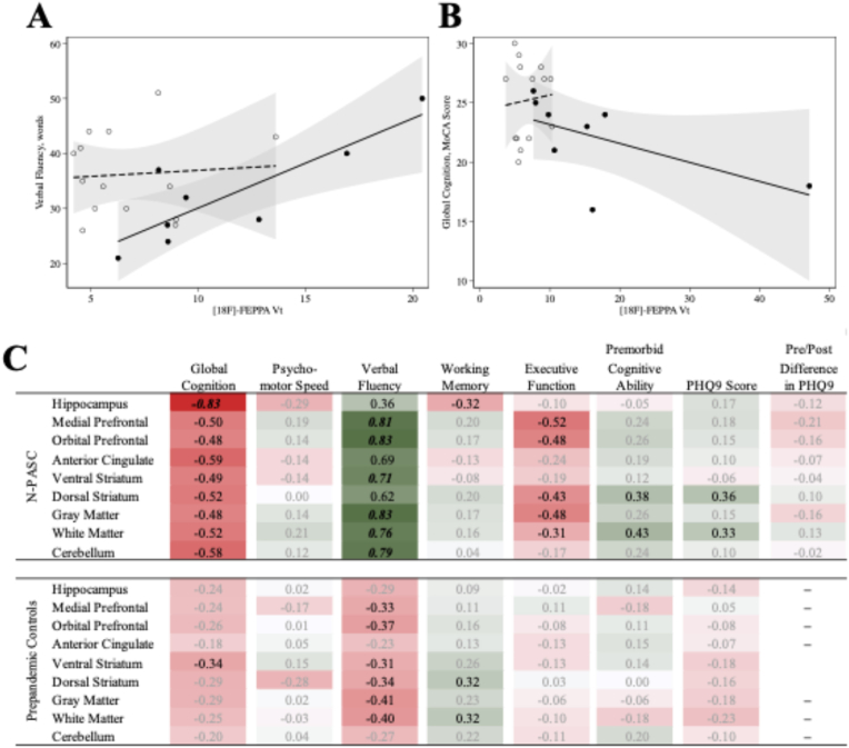
Results: Participants at midlife survived mild to moderate COVID-19 without hospitalization but reported onset of post-acute sequelae of COVID-19 (PASC) for, on average, 22 months before undergoing neuroimaging. Hippocampal VT was higher (VT = 1.70, 95% C.I. = [1.30-2.21], p = 0.001) in participants with persistent brain fog after COVID-19, reflecting an increase of 10.58 mL/cm3 in VT (area under the receiver-operating curve, AUC = 0.95 [0.85-1.00]). At a cutoff of 10.6, sensitivity/specificity/accuracy were 0.88/0.93/0.91.

Conclusion: The results from this study imply that neuroimmune response is a distinct and identifiable characteristic of brain fog after COVID-19. Results suggest that [18F]-FEPPA could be used to support N-PASC diagnosis.

Keywords: Brain Fog; COVID-19; Essential Workers; FEPPA; Glial activation; Positron emission tomography; Post-acute sequelae of COVID-19; Respiratory infection; Translocator protein.


Conflict of interest statement

The authors declare the following financial interests/personal relationships which may be considered as potential competing interests: Sean Clouston reports financial support was provided by 10.13039/100000049National Institute on Aging. Benjamin Luft reports financial support was provided by 10.13039/100000125National Institute for Occupational Safety and Health. If there are other authors, they declare that they have no known competing financial interests or personal relationships that could have appeared to influence the work reported in this paper.

Figures

Image 1
Graphical abstract
Fig. 1
Fig. 1
Violin Plot showing cortical [18F]-FEPPA distribution volume (Vt) in 22 essential workers, comparing N-PASC versus Pre-Pandemic controls and stratified by cerebral region: A: Anterior Cingulate; B: Receiver Operating Curves for N-PASC when examining [18F]-FEPPA binding in the strongest four cerebral regions; C: Medial Prefrontal Cortex; D: Orbital Prefrontal Cortex; E: Hippocampus; F: Ventral Striatum; G: Cerebellum. H: Gray Matter, H: All p-values shown are statistically significant after adjusting for the false discovery rate (FDR = 0.05).
Fig. 2
Fig. 2
Fused [18F]-FEPPA and Structural Image showing differences by standardized uptake values (SUV) in participants with persistent Brain Fog following COVID-19 infection as compared to pre-pandemic controls. Panel A shows TSPO SUV in N-PASC + participants, mapped to the cerebral surface. Panel B shows [18F]-FEPPA SUV values mapped to the cerebral parenchyma. Panel C shows [18F]-FEPPA SUV values for pre-pandemic controls mapped onto the cerebral surface. Panel D shows similar values for [18F]-FEPPA SUV values among pre-pandemic controls throughout the cerebral parenchyma.
Fig. 3
Fig. 3
Associations between regional [18F]-FEPPA VT and cognitive function and depressive symptoms in participants with and without Neurological Post-Acute Sequelae of COVID-19: A) scatter and overlaid linear plots examining association between Gray Matter [18F]-FEPPA VT and Verbal Fluency, stratified into N-PASC + participants (black dots, solid line) and pre-pandemic controls (empty dots, dashed line); B) scatter and overlaid linear plots examining association between Hippocampal [18F]-FEPPA VT and Global Cognition, stratified into N-PASC + participants (black dots, solid line) and pre-pandemic controls (empty dots, dashed line); C) full correlation results reporting Spearman's rho describing associations between regional [18F]-FEPPA Binding (Vt) with all measures of cognitive function and depressive symptoms among participants with Neurological Post-Acute Sequelae of COVID-19. Results for all reasons shown, results with moderate correlations (||rho||>0.30) are shown in black typeface. Statistically significant (p < 0.05) correlations are shown using bold italics.

Similar articles

References

    1. Babalola, et al. SARS-COV-2 re-infection and incidence of post-acute sequelae of COVID-19 (PASC) among essential workers in New York: A retrospective cohort study. The Lancet Regional Health - Americas. 2025;42:100984. - PMC - PubMed
    1. Benedict R.H., et al. Validity of the Symbol Digit Modalities Test as a cognition performance outcome measure for multiple sclerosis. Multiple Sclerosis Journal. 2017;23(5):721–733. - PMC - PubMed
    1. Benjamini Y., Hochberg Y. Controlling the false discovery rate - a practical and powerful approach to multiple testing. Journal of the Royal Statistical Society Series B-Methodological. 1995;57(1):289–300.
    1. Braga J., et al. Neuroinflammation after COVID-19 with persistent depressive and cognitive symptoms. JAMA Psychiatr. 2023;80(8):787–795. - PMC - PubMed
    1. Cai M., et al. Three-year outcomes of post-acute sequelae of COVID-19. Nat. Med. 2024;30(6):1564–1573. - PMC - PubMed

LinkOut - more resources