. 2025 Jan 3;36(1):102446.
doi: 10.1016/j.omtn.2024.102446. eCollection 2025 Mar 11.

miR-125b differentially impacts mineralization in dexamethasone and calcium-treated human mesenchymal stem cells

Affiliations

miR-125b differentially impacts mineralization in dexamethasone and calcium-treated human mesenchymal stem cells

Virginie Joris et al. Mol Ther Nucleic Acids. .

Abstract

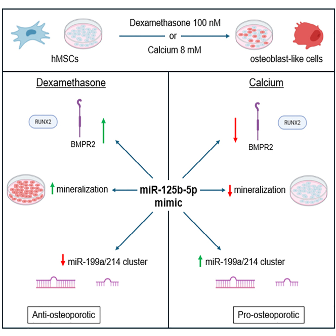
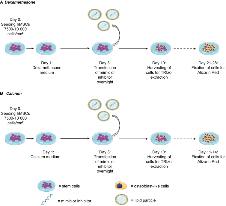
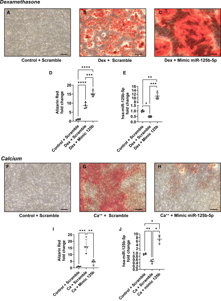
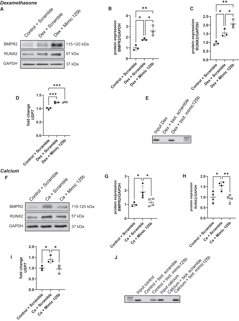
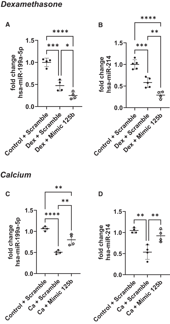
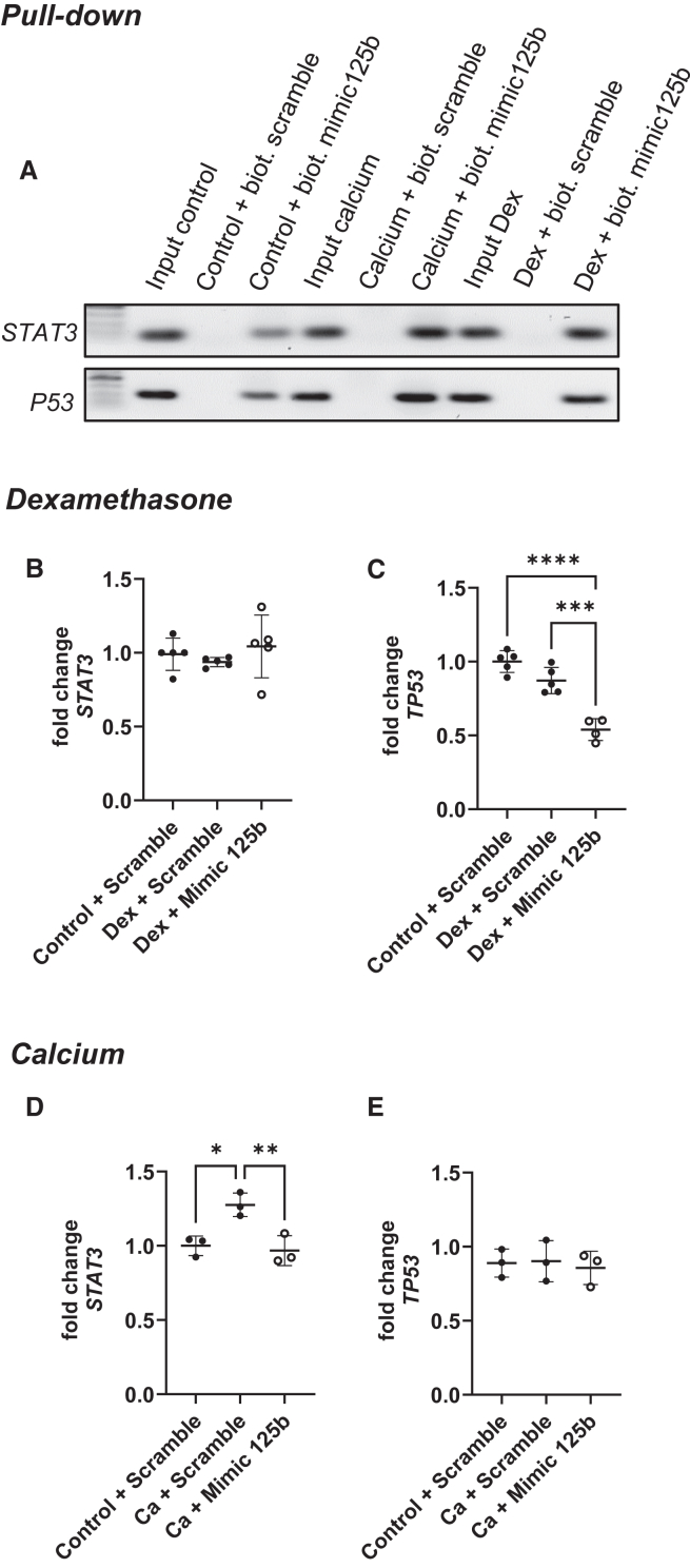
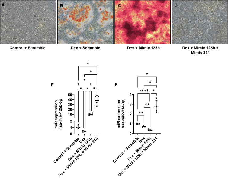
Bone metabolism is highly regulated, and microRNAs (miRs) can contribute to this process. Among them, miR-125b is well known to enhance osteoporosis and reduce osteogenic differentiation of human mesenchymal stem cells (hMSCs). In this work, we aim to evaluate and understand how miR-125b modulates mineralization of hMSCs in two different in vitro models. Cells were cultured in dexamethasone or calcium medium and transfected with miR-125b mimic. Exposure to dexamethasone or calcium medium increased the mineralization of hMSCs and was associated with decreased miR-125b expression. Transfection of miR-125b mimic in dexamethasone-treated cells increased mineralization, while it decreased it in calcium-treated cells. Levels of osteogenic markers presented the same difference. We identified STAT3, p53, and RUNX2 as direct targets of miR-125b in hMSCs. While these targets remained identical in both treatments, their modulation after transfection was different. We showed that miR-125b mimicking differentially modulated the expression of the miR-199a/214 cluster, probably via STAT3/miR-199a/214 and p53/miR-214 pathways. In conclusion, miR-125b affinity for targets implicated in bone remodeling changed depending on the in vitro models used to induce mineralization and led to opposite physiological effects. This work shows the complexity of drugs such as dexamethasone and opens the door for new in vitro models of mineralization.

Keywords: MT: Non-coding RNAs; calcium; dexamethasone; differential effect; miR-125b; mineralization; molecular mechanisms.


Conflict of interest statement

The authors declare no competing interests.

Figures

None
Graphical abstract
Figure 1
Figure 1
Schematic representation of dexamethasone- and calcium-induced mineralization Dexamethasone (A) or calcium chloride (B) was added to the cells one day after seeding. Transfection with mimics or negative control was performed at day 3, and cells were harvested 10 days after the beginning of dexamethasone or calcium treatment. Mineralization was assessed via alizarin red after 21–28 days for dexamethasone and 11–14 days for calcium protocol. Calcium medium, calcium chloride 8 mM and ascorbic acid; dexamethasone medium, dexamethasone 100 nM, β-glycerol phosphate, and ascorbic acid.
Figure 2
Figure 2
Mimicking of miR-125b induced opposite effect in dexamethasone- and calcium-treated hMSCs hMSCs were transfected with negative control or mimic and cultured in control or dexamethasone medium (top) and in control or calcium medium (bottom). Mineralization was assessed with alizarin red (A–C and F–H) and quantified with CPC (D and I) using a spectrophotometer; ∗∗p < 0.01; ∗∗∗p < 0.001; ∗∗∗∗p < 0.0001. miRNA-125b expression was assessed using qPCR (E and J); ∗p < 0.05; ∗∗p < 0.01; ∗∗∗p < 0.001 based on fold change relative to control + scramble. Scale bar, 150 μm. N = 4; n = 2. Results are expressed as mean ± SD.
Figure 3
Figure 3
Mimicking of miR-125b induced the opposite profile of osteogenic proteins level hMSCs were transfected with negative control or mimic and cultured in control or dexamethasone medium (top) and in control or calcium medium (bottom). BMPR2 and RUNX2 levels were measured via western blotting (A and F). The quantification of protein levels was performed using ImageJ normalized to GAPDH (B, C, G, and H); ∗p < 0.05; ∗∗p < 0.01. USP7 expression was assessed using qPCR (D and I); ∗p < 0.05; ∗∗∗p < 0.001 based on fold change relative to control + scramble. The verification of RUNX2 as a direct target of miR-125b was performed using a pull-down assay with biotinylated mimic-125b and negative control combined with PCR and agarose gel (E and J). N = 3–4; n = 2. Results are expressed as mean ± SD.
Figure 4
Figure 4
Mimicking miR-125b differentially regulated miR-199a/214 cluster in dexamethasone- and calcium-treated hMSCs hMSCs were transfected with negative control or mimic and cultured in control or dexamethasone medium (top) and in control or calcium medium (bottom). miR-199a-5p and miR-214 expression was measured using qPCR. ∗p < 0.05; ∗∗p < 0.01; ∗∗∗p < 0.001; ∗∗∗∗p < 0.0001 based on fold change relative to control + scramble. N = 4–5; n = 2. Results are expressed as mean ± SD.
Figure 5
Figure 5
The direct targets STAT3 and p53 reacted differently to miR-125b in dexamethasone and calcium medium The verification of STAT3 and TP53 as a direct target of miR-125b was performed using a pull-down assay with biotinylated mimic-125b and negative control combined with PCR and agarose gel (A). hMSCs were transfected with negative control or mimic and cultured in control or dexamethasone medium (center) and in control or calcium medium (bottom). STAT3 (B and D) and p53 (C and E) expression was measured using qPCR. ∗p < 0.05; ∗∗p < 0.01; ∗∗∗p < 0.001; ∗∗∗∗p < 0.0001 based on fold change relative to control + scramble. N = 3–5; n = 2. Results are expressed as mean ± SD.
Figure 6
Figure 6
Rescuing miR-214 in dexamethasone-treated hMSCs transfected with miR-125b abrogated mineralization hMSCs were transfected with negative control or mimic and cultured in control or dexamethasone medium. Mineralization was assessed with alizarin red (A–D). miRNA-125b and miR-214 expression was assessed using qPCR (E and F). ∗p < 0.05; ∗∗p < 0.01; ∗∗∗∗p < 0.0001 based on fold change relative to control + scramble. N = 4; n = 2. Scale bar, 150 μm. Results are expressed as mean ± SD.

Similar articles

References

    1. Li H., Xiao Z., Quarles L.D., Li W. Osteoporosis: Mechanism, Molecular Target and Current Status on Drug Development. Curr. Med. Chem. 2021;28:1489–1507. doi: 10.2174/0929867327666200330142432. - DOI - PMC - PubMed
    1. Cummings S.R., San Martin J., McClung M.R., Siris E.S., Eastell R., Reid I.R., Delmas P., Zoog H.B., Austin M., Wang A., et al. Denosumab for prevention of fractures in postmenopausal women with osteoporosis. N. Engl. J. Med. 2009;361:756–765. doi: 10.1056/NEJMoa0809493. - DOI - PubMed
    1. Infante A., Rodríguez C.I. Osteogenesis and aging: lessons from mesenchymal stem cells. Stem Cell Res. Ther. 2018;9:244. doi: 10.1186/s13287-018-0995-x. - DOI - PMC - PubMed
    1. Ponzetti M., Rucci N. Osteoblast Differentiation and Signaling: Established Concepts and Emerging Topics. Int. J. Mol. Sci. 2021;22 doi: 10.3390/ijms22136651. - DOI - PMC - PubMed
    1. Nakashima K., de Crombrugghe B. Transcriptional mechanisms in osteoblast differentiation and bone formation. Trends Genet. 2003;19:458–466. doi: 10.1016/S0168-9525(03)00176-8. - DOI - PubMed

LinkOut - more resources