

Case Reports
doi: 10.1016/j.hrcr.2024.09.003.
eCollection 2024 Dec.
Macro-re-entry atrial flutter involving the caval side of a persistent Eustachian valve
Affiliations
- PMID: 39897670
- PMCID: PMC11781871
- DOI: 10.1016/j.hrcr.2024.09.003
Item in Clipboard
Case Reports
Macro-re-entry atrial flutter involving the caval side of a persistent Eustachian valve
HeartRhythm Case Rep.
.
No abstract available
Keywords: Ablation; Arrhythmia; Atrial flutter; Congenital heart disease; Imaging.
Conflict of interest statement
The authors have no conflicts of interest to disclose.
Figures

A: A 12-lead electrocardiogram demonstrating features of typical counterclockwise atrial flutter (AFL) with underlying right bundle branch block and extreme axis deviation. B: Prospectively gated cardiac computed tomography scan with sagittal (left image) and coronal (right image) planes demonstrating the large, persistent Eustachian valve (EV) with inferior vena cava (IVC) distention and right atrium (RA) dilation. C: Intracardiac recording from a multipolar electrode during the clinical AFL. The ablation catheter was placed on the IVC side of the EV, demonstrating a fractionated signal during the atrial diastolic period (red arrow). D: Ablation and termination of clinical AFL at this site. ASD = atrial septal defect; LA = left atrium.

A: Propagation map with an inferior view demonstrating an early-meets-late signal at the Eustachian valve (EV). B: Left-lateral view of the propagation map demonstrating the early-meets late signal. C: Entrainment on the EV using the ablation catheter demonstrating an in-circuit response during tachycardia; postpacing interval minus tachycardia cycle length (PPI – TCL) equals 17 milliseconds. D: Entrainment from coronary sinus 7,8 (closer to the cavotricuspid isthmus) demonstrating PPI – TCL = 26 milliseconds.
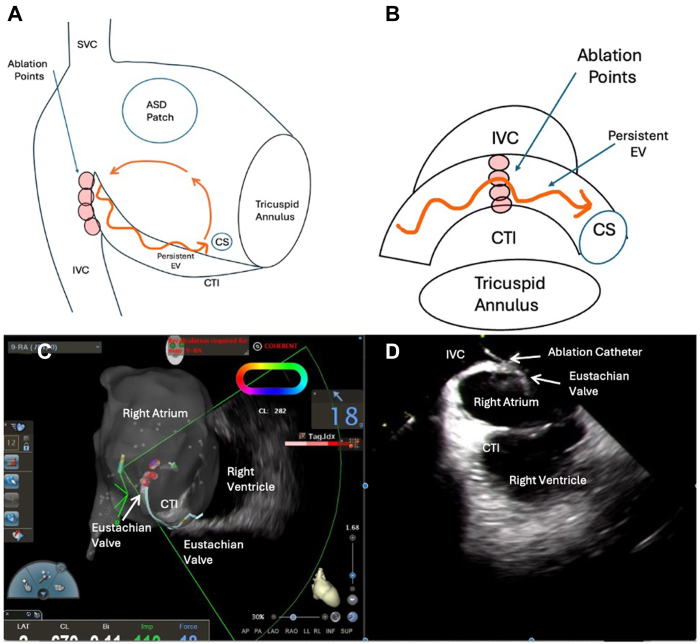
A: Right anterior oblique view demonstrating the circuit based on entrainment and propagation map during atrial flutter. Ablation was delivered on the caval side of the Eustachian valve (EV). B: Focused left anterior oblique view of the same, demonstrating the propagation of the tachycardia across the EV. C: Electroanatomic map of the right atrium in right anterior oblique view. The intracardiac echocardiogram (ICE) catheter fan is directed toward the EV with the ablation catheter on the caval side of the EV. D: Corresponding ICE view from the fan demonstrating the location of the ablation catheter in relation to the EV, with the ablation being performed on the inferior vena cava (IVC) side of the EV. ASD = atrial septal defect; CS = coronary sinus; CTI = cavotricuspid isthmus; SVC = superior vena cava.
References
-
- Casteigt B., Samuel M., Laplante L., et al. Atrial arrhythmias and patient-reported outcomes in adults with congenital heart disease: an international study. Heart Rhythm. 2021;18:793–800. - PubMed
-
- Khairy P., Van Hare G.F., Balaji S., et al. PACES/HRS expert consensus statement on the recognition and management of arrhythmias in adult congenital heart disease: developed in partnership between the Pediatric and Congenital Electrophysiology Society (PACES) and the Heart Rhythm Society (HRS). Endorsed by the governing bodies of PACES, HRS, the American College of Cardiology (ACC), the American Heart Association (AHA), the European Heart Rhythm Association (EHRA), the Canadian Heart Rhythm Society (CHRS), and the International Society for Adult Congenital Heart Disease (ISACHD) Heart Rhythm. 2014;11:e102–e165. - PubMed
-
- Khairy P. EP challenges in adult congenital heart disease. Heart Rhythm. 2008;5:1464–1472. - PubMed
-
- Schuchlenz H.W., Saurer G., Weihs W., Rehak P. Persisting Eustachian valve in adults: relation to patent foramen ovale and cerebrovascular events. J Am Soc Echocardiogr. 2004;17:231–233. - PubMed
-
- Martínez-Quintana E., Rodríguez-González F., Marrero-Santiago H., Santana-Montesdeoca J., López-Gude M.J. Cor triatriatum dexter versus prominent eustachian valve in an adult congenital heart disease patient. Congenit Heart Dis. 2013;8:589–591. - PubMed
Publication types
LinkOut - more resources
Full Text Sources
