

Severe contrast-induced encephalopathy diagnosed with postmortem dual-energy CT in an elderly patient
- PMID: 39897761
- PMCID: PMC11786802
- DOI: 10.1016/j.radcr.2024.12.065
Severe contrast-induced encephalopathy diagnosed with postmortem dual-energy CT in an elderly patient
Abstract
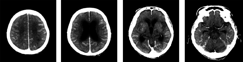
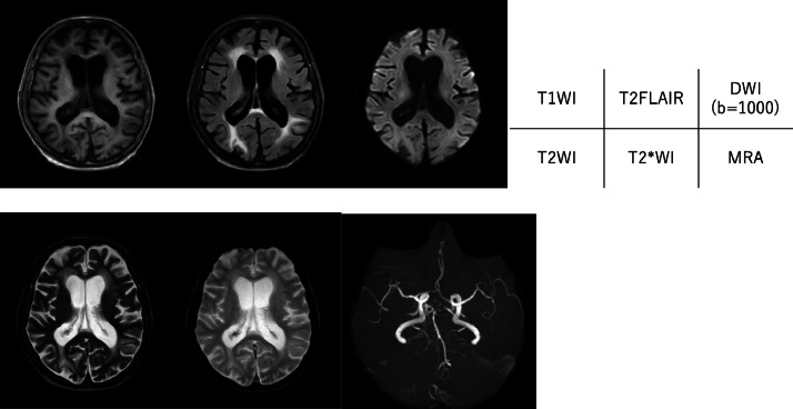
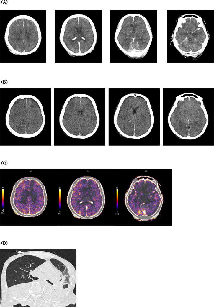
Contrast-induced encephalopathy (CIE) is a rare complication associated with the use of iodine-based contrast agents and can be severe in some cases. In such cases, symptoms of encephalopathy, seizures, and neurological deficits appear shortly after contrast administration. This case report discusses a 90-year-old woman who developed severe CIE after iodine contrast agent administration. The patient underwent contrast enhanced computed tomography (CT) and left lower extremity angioplasty 2 days later. The patient's level of consciousness decreased the day after angioplasty; CT and magnetic resonance imaging (MRI) scans suggested CIE. Although the patient was treated with dialysis, but passed away 2 days after onset. Head CT at the time of onset showed extensive high-density area in the cerebral sulci. However, the distribution was different from typical subarachnoid hemorrhage due to ruptured aneurysm; subsequent MRI showed no evidence of subarachnoid hemorrhage. Therefore, CIE was suspected, rather than hemorrhage. A head dual-energy (DE)-CT, which can non-invasively assess the presence of intracranial iodine, was planned for diagnosing CIE. Although her poor condition made it difficult to performed prior to the death, so postmortem DE-CT was performed and confirmed the presence of iodine intracranially. This case suggests considering CIE in patients who develop impaired consciousness after contrast agents use, even when the contrast agents are not directly injected into cerebral blood vessels. In suspected CIE cases, DE-CT is useful for distinguishing iodine from hemorrhage.
Keywords: Case report; Contrast medium; Contrast-induced encephalopathy; Dual-energy computed tomography; Iodine.
© 2024 The Authors. Published by Elsevier Inc. on behalf of University of Washington.
Figures
References
-
- Kawasaki T., Hayase M., Miyakoshi A., Taki J., Nakamura T., Hatano T. Two cases of symptomatic contrast-induced encephalopathy after coil embolization of unruptured cerebral aneurysm. JNET. 2015;9:96–102. doi: 10.5797/JNET.CR.2015-0002. - DOI
Publication types
LinkOut - more resources
Full Text Sources
