Case Reports
. 2025 Jan 18;20(4):1893-1899.
doi: 10.1016/j.radcr.2024.12.049. eCollection 2025 Apr.

Ischiofemoral impingement syndrome, an unusual entity of hip pain: A case report and literature review

Affiliations
Case Reports

Ischiofemoral impingement syndrome, an unusual entity of hip pain: A case report and literature review

Praveen K Sharma et al. Radiol Case Rep. .

Abstract

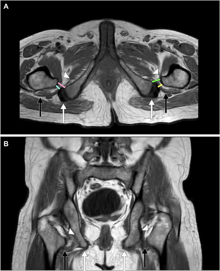
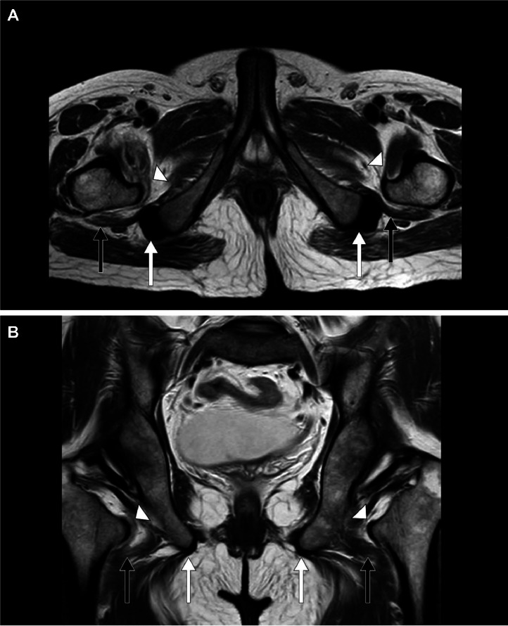
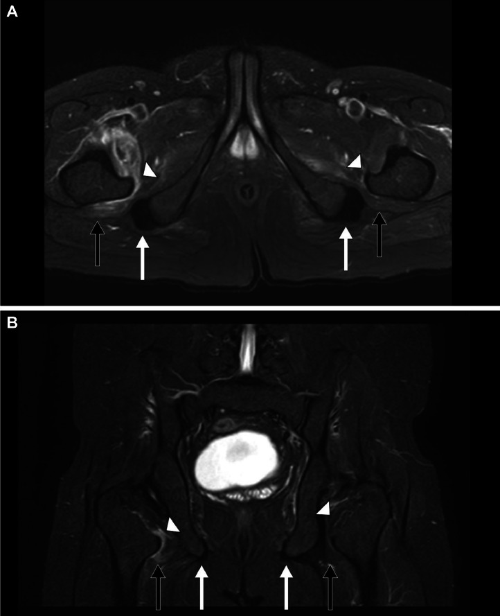
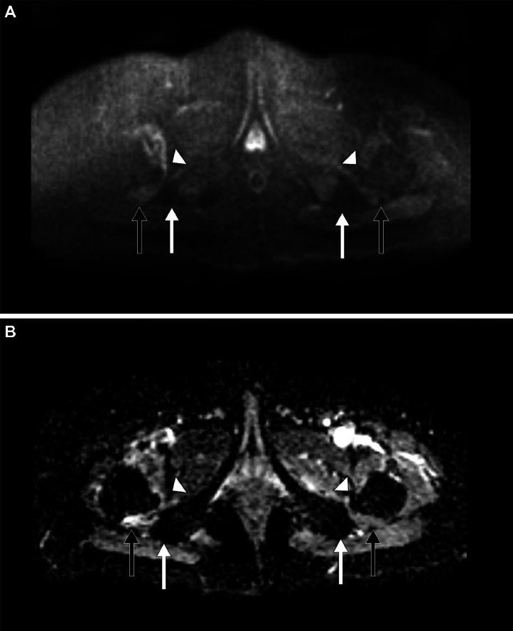
Ischiofemoral impingement syndrome (IFIS) is a rare condition that can cause significant hip pain, often linked to past injuries or surgeries. We present a case of a 33-year-old male who has persistent pain in both hips, radiating down his legs and experiencing a snapping sensation without any history of trauma or surgery. Magnetic resonance imaging (MRI) revealed swelling in the quadratus femoris muscle and reduced space between his ischium and femur, typical signs of IFIS. Instead of opting for surgery, the 33-year-old male managed with anti-inflammatory medications, physical therapy, and a targeted exercise program. The pain gradually subsided, and the 33-year-old male regained complete movement in the hip. This case is noteworthy because it shows that non-surgical treatments can successfully manage IFIS, even in the absence of trauma. This case emphasizes the need to consider IFIS when diagnosing unexplained hip pain.

Keywords: Anti-inflammatory agents; Arthralgia; Exercise therapy; Femur; Ischium; Magnetic resonance imaging.


Figures

Fig 1:
Fig. 1
A 33-year-old male presented to the emergency room (ER) with bilateral progressive hip pain for 2 years, which was insidious in onset and persistent even at rest. Magnetic resonance imaging (MRI) of the hips: T1-weighted (T1W) (A) Axial and (B) Coronal sections shows a mild decrease in the right ischiofemoral space and quadratus femoris space, measuring 11 mm and 10 mm in the distance respectively and a mild decrease in the left ischiofemoral space and quadratus femoris space, measuring 12 mm and 10 mm in the distance respectively with iliopsoas tendons (white arrow heads), quadratus femoris muscles (short black arrows), and hamstring tendons (short white arrows). Note: right ischiofemoral space (pink line), right quadratus femoris space (cyan line), left ischiofemoral space (green line), and left quadratus femoris space (yellow line).
Fig 2:
Fig. 2
A 33-year-old male presented to the emergency room (ER) with bilateral progressive hip pain for 2 years, which was insidious in onset and persistent even at rest. Magnetic resonance imaging (MRI) of the hips: T2-weighted (T2W) (A) Axial and (B) Coronal sections shows a mild decrease in the right ischiofemoral space and quadratus femoris space, measuring 11 mm and 10 mm in the distance respectively and a mild decrease in the left ischiofemoral space and quadratus femoris space, measuring 12 mm and 10 mm in the distance respectively with iliopsoas tendons (white arrow heads), quadratus femoris muscles (short black arrows), and hamstring tendons (short white arrows).
Fig 3:
Fig. 3
A 33-year-old male presented to the emergency room (ER) with bilateral progressive hip pain for 2 years, which was insidious in onset and persistent even at rest. Magnetic resonance imaging (MRI) of the hips: Short tau inversion recovery (STIR) (A) Axial and (B) Coronal sections shows hyperintensities (edema) in the bilateral iliopsoas muscles (white arrow heads), bilateral quadratus femoris muscles (short black arrows), and normal bilateral hamstring tendons (short white arrows).
Fig 4:
Fig. 4
A 33-year-old male presented to the emergency room (ER) with bilateral progressive hip pain for 2 years, which was insidious in onset and persistent even at rest. Magnetic resonance imaging (MRI) of the hips: (A) Diffusion-weighted imaging (DWI) Axial – no reduced diffusivity, (B) Apparent diffusion coefficient (ADC) Axial – no altered signal intensities)} in the bilateral iliopsoas muscles (white arrow heads), bilateral quadratus femoris muscles (short black arrows), and normal bilateral hamstring tendons (short white arrows).

Similar articles

References

    1. Wu W.T., Chang K.V., Mezian K., Naňka O., Ricci V., Chang H.C., et al. Ischiofemoral impingement syndrome: cclinical and imaging/guidance issues with special focus on ultrasonography. Diagnostics (Basel) 2022;13(1):139. doi: 10.3390/diagnostics13010139. - DOI - PMC - PubMed
    1. Torriani M., Souto S.C., Thomas B.J., Ouellette H., Bredella M.A. Ischiofemoral impingement syndrome: an entity with hip pain and abnormalities of the quadratus femoris muscle. AJR Am J Roentgenol. 2009;193(1):186–190. doi: 10.2214/AJR.08.2090. - DOI - PubMed
    1. Johnson K.A. Impingement of the lesser trochanter on the ischial ramus after total hip arthroplasty: report of three cases. J Bone Joint Surg Am. 1977;59(2):268–269. https://pubmed.ncbi.nlm.nih.gov/845219/ - PubMed
    1. Patti J.W., Ouellette H., Bredella M.A., Torriani M. Impingement of lesser trochanter on ischium as a potential cause for hip pain. Skeletal Radiol. 2008;37(11):939–941. doi: 10.1007/s00256-008-0551-3. - DOI - PubMed
    1. Kassarjian A. Signal abnormalities in the quadratus femoris muscle: tear or impingement? AJR Am J Roentgenol. 2008;190(4):W379. doi: 10.2214/AJR.07.3540. - DOI - PubMed

Publication types

LinkOut - more resources