. 2025 Jan 13;11(2):e41935.
doi: 10.1016/j.heliyon.2025.e41935. eCollection 2025 Jan 30.

ISCOM-type matrix from beta-escin and glycyrrhizin saponins

Affiliations

ISCOM-type matrix from beta-escin and glycyrrhizin saponins

V Petkov et al. Heliyon. .

Abstract

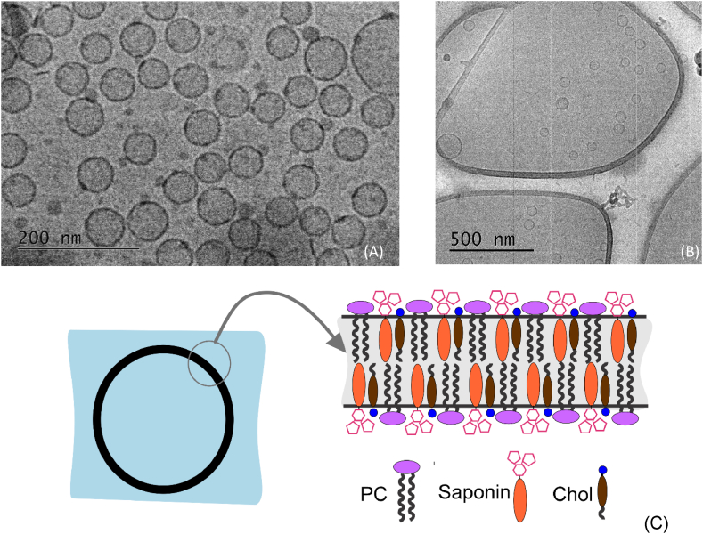
Background and aims: Nanotechnology provides the opportunity for construction of modern transport devices such as nanoparticles for a variety of applications in the field of medicine. A novel experimental protocol for the formation of saponin-cholesterol-phospholipid nanoparticles of vesicular structure has been developed and applied to prepare stable nanoparticles using escin or glycyrrhizin as saponins.

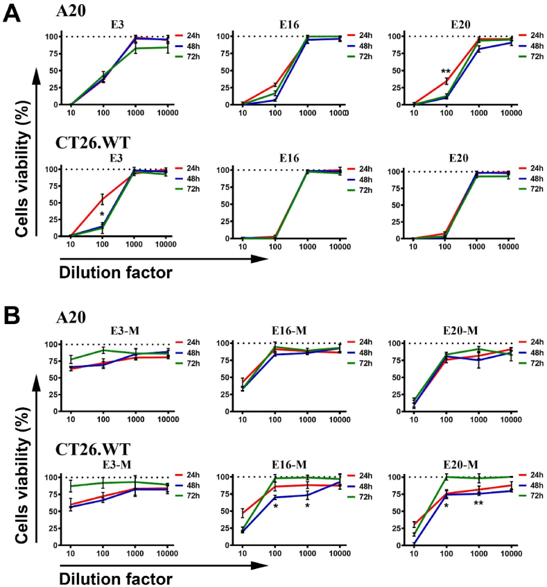
Methods: The methods for nanoparticle construction include a sonication at 90 °C of the initial mixture of components, followed by an additional sonication on the next day for incorporation of an additional amount of cholesterol, thus forming stable unilamellar vesicles. Tests and assays for cell viability, erythrocyte hemolysis, flow cytometry, and fluorescent microscopy analyses have been performed.

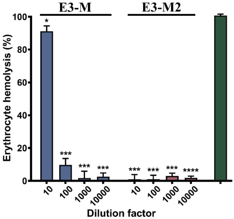
Results: By selecting appropriate component ratios, stable and safe particles were formulated with respect to the tested bio-cells. The prepared nanoparticles have mean diameter between 70 and 130 nm, depending on their composition. The versatility of these nanoparticles allows for the encapsulation of various molecules, either within the vesicle interior for water-soluble components or within the vesicle walls for hydrophobic components. The saponin particles formed after cholesterol post-addition (E3-M2) are stable and 100 % of the cells remain viable even after 10-times dilution of the initial particle suspension. These particles are successful included into isolated mouse macrophages.

Conclusions: Among the variety of generated nanoparticles, the E3-M2 particles demonstrated properties of safe and efficient devices for future vaccine design and antigen targeting to immune system.

Keywords: Immune targeting device; Liposomes; Nanoparticles; Toxicity.


Conflict of interest statement

The authors declare that they have no known competing financial interests or personal relationships that could have appeared to influence the work reported in this paper.

Figures

Image 1
Graphical abstract
Fig. 1
Fig. 1
A20 and CT26.WT cells viability after 24 h (red curves), 48 h (blue curves), and 72 h (green curves) after treatments with particles as a function of dilution. Untreated cells were used as controls with 100 % viability for each time period. 1A. Treatment of cell lines with samples E3, E16 and E20 from Table 1; 1B. The same treatment of cell lines with modified particles (samples E3-M, E16-M and E20-M from Table 2). All samples were triplicated and mean ± SD values were presented for each group; p values were calculated using the two-way ANOVA test (∗p < 0.05; ∗∗p < 0.01). A representative of three independent experiments is shown.
Fig. 2
Fig. 2
Cryo-TEM images of the particles from E3-M2 sample, 200 keV, magnification 40000×(A) Without fluorescence dye; (B) after incorporation of Nile red, magnification 10 000×; (C) Schematic presentation of the ISCOM particles formed in the current study. The composition of the bilayer of the formed particles is schematically shown: Saponin = escin; Chol = cholesterol; PC = 1,2-dipalmitoyl-sn-glycero-3-phosphocholine.
Fig. 3
Fig. 3
A20 and CT26.WT cell viability after 24 h (red curves); 48 h (blue curves) and 72 h (green curves) after treatments with modified particles from samples E3-M; E3-M2; G3-M and G3-M2 (from Table 2) as a function of dilution. Untreated cells were used as controls with 100 % viability for each time period. All samples were triplicated and mean ± SD values were presented for each group; p values were calculated using the two-way ANOVA test. A representative of three independent experiments is shown.
Fig. 4
Fig. 4
Erythrocyte hemolysis induced by E3-M and E3-M2 particles as a function of dilution. All samples were triplicated and mean ± SD values were presented for each group; p values were calculated using the two-way ANOVA test (∗p < 0.05; ∗∗∗p < 0.001; ∗∗∗∗p < 0.0001) compared to 100 % hemolysis control. A representative of three independent experiments is shown.
Fig. 5
Fig. 5
(A) FACS assay for the integration of the E3-M2-Nile Red nanoparticles into the F4/80-positive cells. Isolated mouse macrophages were co-cultured with the nanoparticles E3-M2-Nile Red, and the staining with F4/80-FITC and E3-M2-Nile Red incorporation were measured by flow cytometry. The percentage of stained cells is shown in the quadrants. A representative of four independent experiments is shown. (B) Fluorescence microscopy analysis of the E3-M2-Nile Red included nanoparticles combined with staining of nuclei and actin (bars = 20 μm, magnification 20×). Control cells without E3-M2-Nile Red particles (Line A) validated the protocol and the initial shape of the cells. Experimental slides with E3-M2-Nile Red particles, (Lines B, C, D, E, F) showed the integration of the particles within the cells. Actin Green (Column 1) stained the actin in the cells, while Hoechst stain (Column 2) was used to colour the cells nuclei. The Nile Red E3-M2 included particles (column 3) were observed in the red spectrum. Merged images of all columns are represented in Column 4.

References

    1. Morein B., Sundquist B., Hoglund S., Dalsgaard K., Osterhaus A. Iscom, a novel structure for antigenic presentation of membrane proteins from enveloped viruses. Nature. 1984;308:457–460. - PubMed
    1. Peek L.J., Russell Middaugh C., Berkland Cory. Nanotechnology in vaccine delivery. Adv. Drug Deliv. Rev. 2008;60(8):915–928. doi: 10.1016/j.addr.2007.05.017. - DOI - PMC - PubMed
    1. Sharma S., Mukkur T., Benson H.A., Chen Y. Pharmaceutical aspects of intranasal delivery of vaccines using particulate systems. J. Pharmaceut. Sci. 2009;98:812–843. - PubMed
    1. Zhao L., Seth A., Wibowo N., Zhao C.X., Mitter N., Yu C., Middelberg A.P. Nanoparticle vaccines. Vaccine. 2014;32(3):327–337. doi: 10.1016/j.vaccine.2013.11.069. PMID: 24295808. - DOI - PubMed
    1. Pearse M.J., Drane D. ISCOMATRIXTM adjuvant: a potent inducer of humoral and cellular immune responses. Vaccine. 2004;22:2391–2395. - PubMed

LinkOut - more resources