

Topical transdermal administration of lenalidomide nanosuspensions-based hydrogels against melanoma: In vitro and in vivo studies
- PMID: 39898009
- PMCID: PMC11787432
- DOI: 10.1016/j.ijpx.2025.100316
Topical transdermal administration of lenalidomide nanosuspensions-based hydrogels against melanoma: In vitro and in vivo studies
Abstract
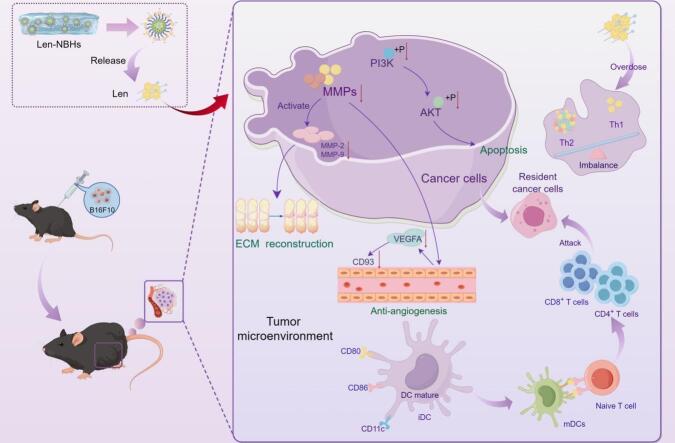
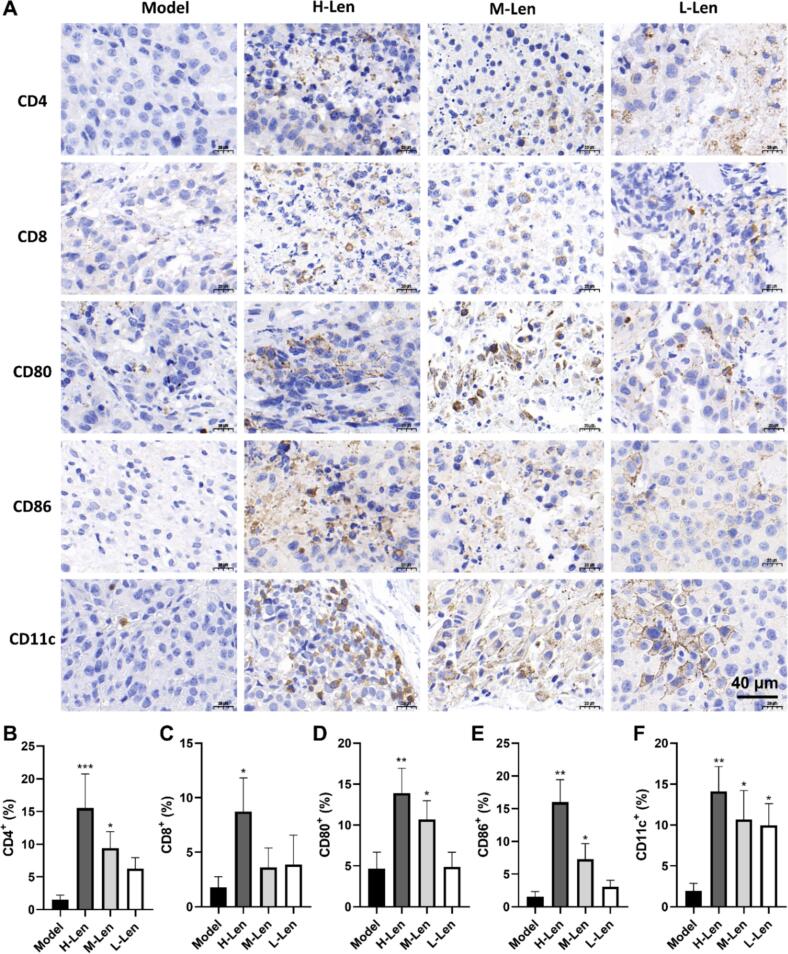
Percutaneous neoadjuvant therapy has proven effective in diminishing tumor size and the surgical intervention area, which couldeffectively mitigate the risk of tumor recurrence and enhance immunotherapy efficacy. Lenalidomide, an approved medication orally used to treat myeloma, was loaded into nanosuspensions-based hydrogels (Len-NBHs) for transdermal administration as a percutaneous neoadjuvant therapy. This study was designed to investigate the inhibitory effect and mechanism of Len-NBHs on melanoma. Network pharmacology and transcriptomic analyses identified key targets and signaling pathways. The effects of lenalidomide on melanoma were further verified through Western blotting, immunohistochemistry, immunofluorescence, and quantitative real-time polymerase chain reaction,using both in vitro cell experiments and in vivo melanoma mouse models. Lenalidomide could induce melanoma cells apoptosis, disrupt cell cycle progression, impede cell migration and invasion, and modify tumor microenvironment (TME). Mechanistically, lenalidomide reversed the abnormal activation of the PI3K-AKT signaling pathway and the overexpression of CD93, while also recruiting CD8+ T cells, CD4+ T cells, and dendritic cells to infiltrate the tumor site. Transdermal administration of Len-NBHs represents a promising adjuvant therapy for the treatment of malignant melanoma. Preoperative administration of Len-NBHs can inhibit the outward spread of melanoma, reduce tumor size, thereby decreasing the surgical excision area and improving patient survival rates and prognosis.
Keywords: Immunoregulation; Lenalidomide nanosuspensions-based hydrogels; Melanoma Transcriptomic; Transdermal administration.
© 2025 The Authors.
Conflict of interest statement
The authors declare that they have no known competing financial interests or personal relationships that could have appeared to influence the work reported in this paper.
Figures


Similar articles
-
Lenalidomide attenuates IMQ-induced inflammation in a mouse model of psoriasis.Biomed Pharmacother. 2022 Dec;156:113883. doi: 10.1016/j.biopha.2022.113883. Epub 2022 Oct 18. Biomed Pharmacother. 2022. PMID: 36270258
-
Lenalidomide overcomes the resistance to third-generation CD19-CAR-T cell therapy in preclinical models of diffuse large B-cell lymphoma.Cell Oncol (Dordr). 2023 Aug;46(4):1143-1157. doi: 10.1007/s13402-023-00833-6. Epub 2023 May 23. Cell Oncol (Dordr). 2023. PMID: 37219767
-
Lenalidomide enhances antitumor functions of chimeric antigen receptor modified T cells.Oncoimmunology. 2015 Dec 3;5(4):e1115940. doi: 10.1080/2162402X.2015.1115940. eCollection 2016 Apr. Oncoimmunology. 2015. PMID: 27141398 Free PMC article.
-
Prognostic indicators of lenalidomide for multiple myeloma: consensus and controversy.Expert Rev Anticancer Ther. 2015;15(7):787-804. doi: 10.1586/14737140.2015.1044249. Epub 2015 May 6. Expert Rev Anticancer Ther. 2015. PMID: 25947283 Review.
-
How lenalidomide is changing the treatment of patients with multiple myeloma.Crit Rev Oncol Hematol. 2013 Oct;88 Suppl 1:S23-35. doi: 10.1016/j.critrevonc.2013.05.013. Epub 2013 Jun 28. Crit Rev Oncol Hematol. 2013. PMID: 23816163 Review.
References
-
- Aue G., Njuguna N., Tian X., Soto S., Hughes T., Vire B., Keyvanfar K., Gibellini F., Valdez J., Boss C., Samsel L., McCoy J.P., Jr., Wilson W.H., Pittaluga S., Wiestner A. Lenalidomide-induced upregulation of CD80 on tumor cells correlates with T-cell activation, the rapid onset of a cytokine release syndrome and leukemic cell clearance in chronic lymphocytic leukemia. Haematologica. 2009;94:1266–1273. - PMC - PubMed
-
- Backlund E., Grozman V., Egyhazi Brage S., Lewensohn R., Lindberg K., Helgadottir H. Radiotherapy with or without immunotherapy in metastatic melanoma: efficacy and tolerability. Acta Oncol. 2023;62:1921–1930. - PubMed
-
- Bertazza L., Mocellin S. The dual role of tumor necrosis factor (TNF) in cancer biology. Curr. Med. Chem. 2010;17:3337–3352. - PubMed
LinkOut - more resources
Full Text Sources
Other Literature Sources
Research Materials
Miscellaneous
