. 2024 Dec 18;28(2):111632.
doi: 10.1016/j.isci.2024.111632. eCollection 2025 Feb 21.

MERS-CoV spike vaccine-induced N-terminal domain-specific antibodies are more protective than receptor binding domain-specific antibodies

Affiliations

MERS-CoV spike vaccine-induced N-terminal domain-specific antibodies are more protective than receptor binding domain-specific antibodies

Olubukola M Abiona et al. iScience. .

Abstract

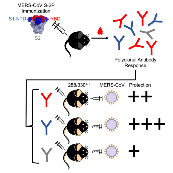
The COVID-19 pandemic underscores the need to prepare for future emerging coronavriuses (CoVs) by understanding the principles behind effective CoV vaccine design such as protective immunity and antibody responses. To study which epitopes and subdomains contribute to in vivo protection, we utilized the prefusion-stabilized spike protein of MERS-CoV, MERS S-2P, as a vaccine immunogen. Vaccination with MERS S-2P elicited both receptor-binding domain (RBD)- and non-RBD-specific antibodies, including N-terminal domain (NTD)-specific G2-and CDC2-A2-like antibodies. Intriguingly, the immunogen MERS S-2P_ΔRBD, MERS S-2P with the RBDs removed, protects comparably to S1 and S-2P immunogens against MERS-CoV challenge. Moreover, passive transfer studies of polyclonal IgG from MERS S-2P immunized mice depleted of subdomain-specific antibodies demonstrated that non-RBD antibodies protected more than non-NTD antibodies. Altogether, these findings illustrate that in-vivo protection is not solely driven by RBD-specific antibodies and highlights the importance of targeting non-RBD sites in future CoV vaccine designs.

Keywords: Immunity; Immunology; Virology.


Conflict of interest statement

N.W., K.S.C-H, M.K., A.W., B.S.G., and J.S.M. are inventors on a US patent entitled “Prefusion Coronavirus Spike Proteins and Their Use.” L.W., W.S., W.-P.K., M.K., J.R.M., and B.S.G. are inventors on a US patent application entitled “Middle East Respiratory Syndrome Coronavirus Immunogens, Antibodies, and Their Use.”

Figures

None
Graphical abstract
Figure 1
Figure 1
Dissection of MERS S domain-specific antibody responses (A–F) Sera from MERS S1 (orange), S-2P (red), and S-2P_ΔRBD (purple) immunized mice (n = 10/group) were pooled and depleted of specificity to non-specific trimeric viral fusion protein (white), MERS S-2P (dots), MERS S1 (horizontal lines), MERS NTD (diagonal lines), or MERS RBD (vertical lines), respectively and assessed for (A-C) binding to MERS S-2P protein by fold-on-competed ELISA and (D-F) neutralizing antibodies against MERS M35c4 pseudovirus.
Figure 2
Figure 2
Ability of prefusion-stabilized MERS S trimers to protect mice from lethal challenge (A–F) 288/330+/+ mice were immunized at week(s) 0 and 3 with PBS (black), MERS S1 (orange), MERS S-2P (red), or MERS S-2P_ΔRBD (purple) and challenged 4 weeks post-boost with a lethal dose of MERS-CoV. Experimental groups were compared by 1-way ANOVA with the Kruskal–Wallis post-test. ∗ = p-value <0.05, ∗∗ = p-value <0.01, ∗∗∗ = p-value <0.001, ∗∗∗∗ = p-value <0.0001. (A) Pre-challenge sera were collected from a subset of mice and assessed for neutralizing antibodies against MERS m35c4 pseudovirus. The dotted line represents the assay limit of detection. Each symbol represents an individual mouse. Bars represent the geometric mean titer (GMT) of each group. (B) Following challenge, mice were monitored for weight loss. The mean of each group is represented by symbols. Error bars represent SEMs. The dotted line represents the threshold of weight loss at which mice were sacrificed. (C–F) At days 3 (C,E) and 5 (D,F) post-challenge, a subset of mouse lungs was harvested for analysis of (C-D) congestion score (0 = no discoloration, 4 = 100% discoloration in all lobes) and (E-F) viral titers. Each symbol represents an individual mouse. Bars represent the (C-D) mean or (E-F) GMT of each group. Error bars represent (C-D) SEMs or (E-F) geometric standard deviations (SDs).
Figure 3
Figure 3
Ability of MERS S domain-specific IgGs to protect mice from lethal challenge (A–E) IgGs were purified from MERS S-2P immunized wild-type mice, depleted of specificity to non-specific trimeric viral fusion protein (black), MERS S-2P (red), MERS S1 (orange), MERS NTD (purple), or MERS RBD (green) and passively transferred to 288/330+/+ mice. At 24 h post-immunization, mice were challenged with a lethal dose of MERS-CoV m35c4. (A) Following challenge, mice were monitored for weight loss. The mean of each group is represented by symbols. Error bars represent SEMs. The dotted line represents the threshold of weight loss at which mice were sacrificed. Experimental groups were compared to the mock-depleted group by 1-way ANOVA with the Kruskal–Wallis post-test. ∗ = p-value <0.05, ∗∗ = p-value <0.01, ∗∗∗ = p-value <0.001. Weight loss curves were completed in two separate experiments with similar results. (B–E) At 3 and 5 days post-challenge, a subset of mouse lungs was harvested for analysis of (B-C) congestion score (0 = no discoloration, 4 = 100% discoloration in all lobes) and (D-E) viral titer. Each symbol represents an individual mouse. Bars represent the (B-C) mean or (D-E) GMT of each group. Error bars represent (B-C) SEMs or (D-E) geometric SDs. Groups were compared by 1-way ANOVA with the Kruskal–Wallis post-test. ∗ = p-value <0.05, ∗∗ = p-value <0.01.
Figure 4
Figure 4
MERS S-2P trimer vaccination elicits G2-and CDC-A2-like NTD-specific Abs (A) Representative 2D class averages from negative-stain EM analysis of MERS S-2P complexed with Fabs digested from RBD-depleted serum polyclonal IgGs. (B) Refined 3D reconstructions from negative-stain EM analysis of MERS S-2P complexed with Fabs digested from RBD-depleted serum polyclonal IgGs. (C) Side and top view of a composite 3D figure demonstrating the two NTD Fab specificities in purple and orange. (D) Side and top views of composite 3D figures comparing each NTD specificity from polyclonal analysis (shown as mesh) to monoclonal antibodies G2 (solid purple) or CDC2-A2 (solid orange). (E) Mesh representation of the composite figure shown in (C) with MERS spike structure (PDB# 5X5F) docked into the density for epitope depiction.
Figure 5
Figure 5
Protective antibody thresholds of RBD- and NTD-specific mAbs (A and B) 288/330+/+ mice were passively immunized with 1 mg/kg (closed squares) or 0.2 mg/mg (open squares) of RBD-specific mAb, CDC-C2 (green), G2 (purple), or an isotype control (black). At 12 h post-immunization, mice were challenged with a lethal dose of MERS-CoV m35c4. Following challenge, mice were monitored for weight loss. The mean of each group is represented by symbols. Error bars represent standard errors of the mean (SEMs). Doses were compared by 1-way ANOVA with the Kruskal–Wallis post-test. ∗ = p-value <0.05, ∗∗∗ = p-value <0.001. (C and D) 288/330+/+ mice were bled 11 h following immunization and serum was assessed for the (C) quantity of MERS S-2P-specific binding antibodies or (D) reciprocal IC50 titer of serum neutralizing antibodies. Each symbol represents an individual mouse. Box represents the interquartile range and whiskers depict minimum and maximum. Groups were compared by the Mann–Whitney t-test. ∗ = p-value <0.05, ∗∗ = p-value <0.01.

References

    1. Fan Y., Zhao K., Shi Z.L., Zhou P. Bat Coronaviruses in China. Viruses. 2019;11 doi: 10.3390/v11030210. - DOI - PMC - PubMed
    1. Zumla A., Peiris M., Memish Z.A., Perlman S. Anticipating a MERS-like coronavirus as a potential pandemic threat. Lancet. 2024;403:1729–1731. doi: 10.1016/S0140-6736(24)00641-X. - DOI - PubMed
    1. Singh S.K. Middle East Respiratory Syndrome Virus Pathogenesis. Semin. Respir. Crit. Care Med. 2016;37:572–577. doi: 10.1055/s-0036-1584796. - DOI - PMC - PubMed
    1. Widagdo W., Sooksawasdi Na Ayudhya S., Hundie G.B., Haagmans B.L. Host Determinants of MERS-CoV Transmission and Pathogenesis. Viruses. 2019;11:280. doi: 10.3390/v11030280. - DOI - PMC - PubMed
    1. Chen Y., Lu S., Jia H., Deng Y., Zhou J., Huang B., Yu Y., Lan J., Wang W., Lou Y., et al. A novel neutralizing monoclonal antibody targeting the N-terminal domain of the MERS-CoV spike protein. Emerg. Microb. Infect. 2017;6:e37. doi: 10.1038/emi.2017.18. - DOI - PMC - PubMed

LinkOut - more resources