. 2025 Jan 2;28(2):111726.
doi: 10.1016/j.isci.2024.111726. eCollection 2025 Feb 21.

Snapshots of Pseudomonas aeruginosa SOS response reveal structural requisites for LexA autoproteolysis

Affiliations

Snapshots of Pseudomonas aeruginosa SOS response reveal structural requisites for LexA autoproteolysis

Filippo Vascon et al. iScience. .

Abstract

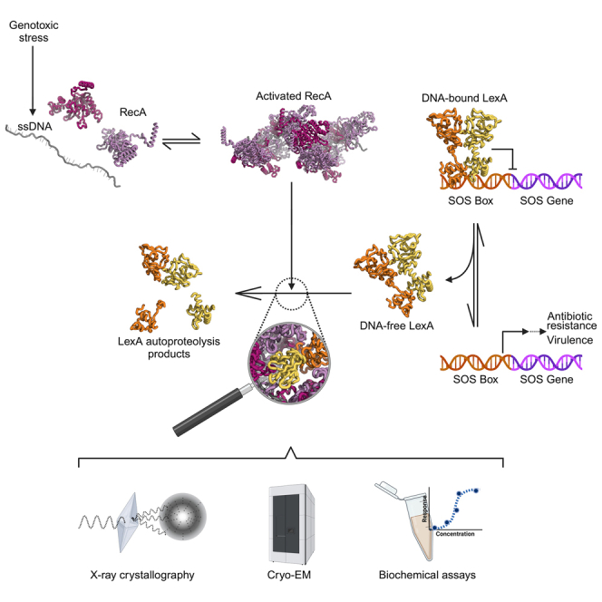
Antimicrobial resistance poses a severe threat to human health and Pseudomonas aeruginosa stands out among the pathogens responsible for this emergency. The SOS response to DNA damage is crucial in bacterial evolution, influencing resistance development and adaptability in challenging environments, especially under antibiotic exposure. Recombinase A (RecA) and the transcriptional repressor LexA are the key players that orchestrate this process, determining either the silencing or the active transcription of the genes under their control. By integrating state-of-the-art structural approaches with in vitro binding and functional assays, we elucidated the molecular events activating the SOS response in P. aeruginosa, focusing on the RecA-LexA interaction. Our findings identify the conserved determinants and strength of the interactions that allow RecA to trigger LexA autocleavage and inactivation. These results provide the groundwork for designing novel antimicrobial strategies and exploring the potential translation of Escherichia coli-derived approaches, to address the implications of P. aeruginosa infections.

Keywords: Biological sciences; Biophysics; Microbiology; Natural sciences.


Conflict of interest statement

The authors declare no competing interests.

Figures

None
Graphical abstract
Figure 1
Figure 1
Structural analysis of LexAPaCTD (A) Analytical size exclusion chromatography of LexAPaS125A (blue) and LexAPaCTDG91D (yellow; chromatograms on the left and standard curve interpolation on the right). (B) SDS-PAGE-based RecAPa∗-induced autoproteolysis assay of 4 LexAPa variants: full-length LexAPa, either wt or S125A inactive mutant, and LexAPaCTD, either wt or G91D uncleavable mutant. One representative gel is shown, see also Figure S1E for band quantification. (C) Overall view of the LexAPaCTDG91D dimer (chains A and B), as revealed by X-ray crystallography. The catalytic dyad (S125/K162) and the mutated self-cleavage site (A90-D91) of each monomer are shown as orange sticks. Boxed regions are zoomed in panels D and E. Superposed (transparent green cartoon) is the closed conformation of LexAPa cleavable loop found in LexAPaS125A bound to RecAPa∗. (D) Detailed view of the cleavable loop (chain A) in the “open” (inactive) conformation. Hydrogen bonds engaging the residues of the loop are represented as dashed lines, while residues involved in a hydrophobic cluster are depicted as orange sticks. (E) Detailed views of the homodimerization surface of LexAPaCTD. Dashed lines indicate H-bonds, salt bridges and cation-π interactions, while residues involved in a hydrophobic cluster are depicted as orange sticks. (F) LexAPa cleavable loop in the “closed” (active) conformation. Dashed lines indicate H-bonds stabilizing the loop in this state, while orange sticks correspond to the catalytic dyad and to the hydrophobic residues indicated in panel B. The movement of the loop brings the cleavage site inside the catalytic pocket and at the same time opens a hydrophobic cavity (Y117, L119, V159, I85) that hosts I94 in the open conformation.
Figure 2
Figure 2
Cryo-EM structure of RecAPa∗ (A) RecAPa∗ cryo-EM density map. (B–D) Coloring of density regions corresponding to RecAPa∗ protomers and (C and D) zoom on the atomic model (two perpendicular views). (E) Zoom on two adjacent RecAPa∗ protomers assembled on ssDNA (RecAPan and RecAPan+1, moving from 5′ to 3′ on ssDNA). Detailed views of the cryo-EM map around ssDNA (F) and ATPγS (G), and RecAPa residues interacting with them.
Figure 3
Figure 3
Cryo-EM structure of RecAPa∗-LexAPaS125A (A) Cryo-EM density map of the RecAPa-LexAPaS125A complex. The displayed map has been locally sharpened using LocScale2. (B) Coloring of density regions corresponding to RecAPa∗ protomers (purple tones) and LexAPa CTD chains A (yellow) and B (orange). The boxed region represents a low-resolution density, which was not interpreted by the atomic model and that might be due to the LexAPa NTD. (C and D) (C) Side and (D) front views of the RecAPa∗-LexAPaS125A atomic model. The dashed line in panel C represents a virtual plane where the model was cut in panel D to allow LexAPa clear visualization. (E) Electrostatic surface potential of RecAPa∗ and LexAPaCTD, showing complementarity on the interacting surfaces. (F) LexAPaCTD dimer and the main binding determinants on four RecAPa protomers (chains G–J), zoomed in panels (G–J). (K) Details of the interfaces buried between LexAPa and different RecAPa∗ protomers. (L and M) The corresponding interacting surfaces are represented in panels (L) (on RecAPa∗ surface) and (M) (on LexAPa surface, front and side views). Contour lines are colored as the interacting chain.
Figure 4
Figure 4
Analysis of RecAPa interactions with its natural ligands (ATPγS, ssDNA and LexAPa) (A–C) FP-based titrations of (A) FAM-32mer ssDNA with RecAPa (ATPγS in molar excess), (B) RecAPa/FAM-32mer ssDNA with ATPγS and (C) FlAsH-LexAPaCTDS125A with activated RecAPa (RecAPa∗, RecAPa/ssDNA/ATPγS). Points represent the average of three replicates while error bars represent standard errors of the mean (SEM). (D) Relative activity of RecAPa variants (wt, M201A, F202A) in terms of oligomerization on ssDNA and induction of FlAsH-LexAPaCTD autoproteolysis. Bars represent averages of three replicates ±SEM. See also Figure S5 for time-course traces. (E) Overview of the model proposed for the molecular process promoted by RecAPa∗, that leads to the autocleavage of LexAPa. LexAPa can bind RecAPa∗ if it is free from DNA and with the cleavable loop in the closed conformation. The binding to RecAPa∗ allows the self-cleavage of LexAPa, that otherwise is mainly prevented.

Similar articles

References

    1. Murray C.J.L., Ikuta K.S., Sharara F., Swetschinski L., Robles Aguilar G., Gray A., Han C., Bisignano C., Rao P., Wool E., et al. Global burden of bacterial antimicrobial resistance in 2019: a systematic analysis. Lancet. 2022;399:629–655. doi: 10.1016/S0140-6736(21)02724-0. - DOI - PMC - PubMed
    1. Tacconelli E., Carrara E., Savoldi A., Harbarth S., Mendelson M., Monnet D.L., Pulcini C., Kahlmeter G., Kluytmans J., Carmeli Y., et al. Discovery, research, and development of new antibiotics: the WHO priority list of antibiotic-resistant bacteria and tuberculosis. Lancet Infect. Dis. 2018;18:318–327. doi: 10.1016/S1473-3099(17)30753-3. - DOI - PubMed
    1. Pang Z., Raudonis R., Glick B.R., Lin T.-J., Cheng Z. Antibiotic resistance in Pseudomonas aeruginosa: mechanisms and alternative therapeutic strategies. Biotechnol. Adv. 2019;37:177–192. doi: 10.1016/j.biotechadv.2018.11.013. - DOI - PubMed
    1. Rice L.B. Federal Funding for the Study of Antimicrobial Resistance in Nosocomial Pathogens: No ESKAPE. J. Infect. Dis. 2008;197:1079–1081. doi: 10.1086/533452. - DOI - PubMed
    1. Liao C., Huang X., Wang Q., Yao D., Lu W. Virulence Factors of Pseudomonas Aeruginosa and Antivirulence Strategies to Combat Its Drug Resistance. Front. Cell. Infect. Microbiol. 2022;12 doi: 10.3389/fcimb.2022.926758. - DOI - PMC - PubMed

LinkOut - more resources