. 2025 Jan 4;28(2):111752.
doi: 10.1016/j.isci.2025.111752. eCollection 2025 Feb 21.

Extracellular vesicles contribute to the beneficial effects of exercise training in APP/PS1 mice

Affiliations

Extracellular vesicles contribute to the beneficial effects of exercise training in APP/PS1 mice

Oliver K Fuller et al. iScience. .

Abstract

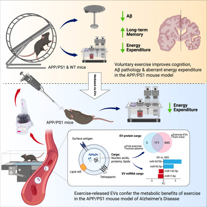
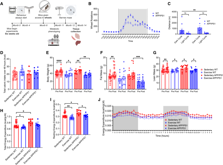
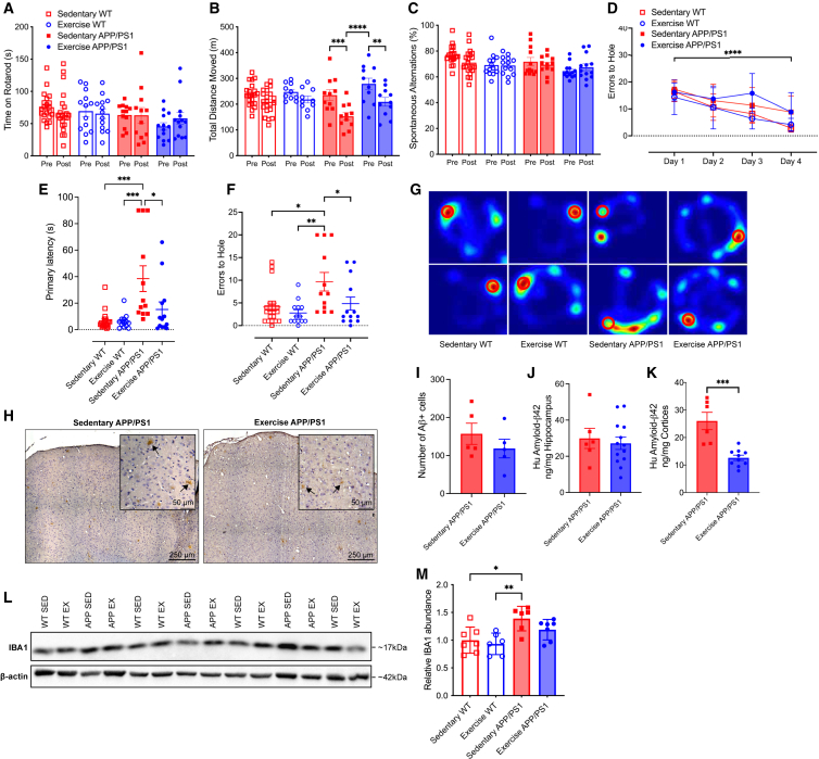
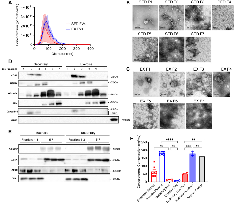
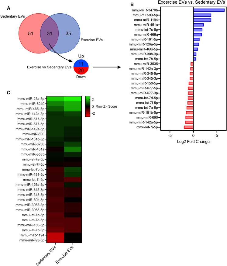
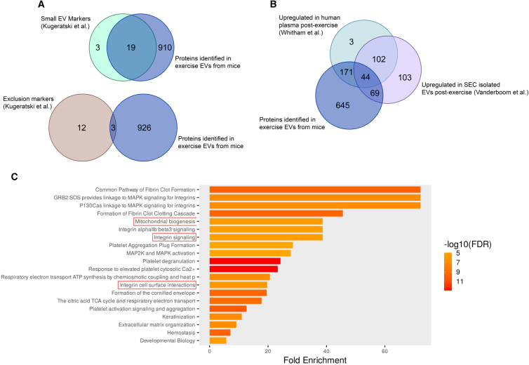
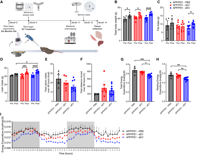
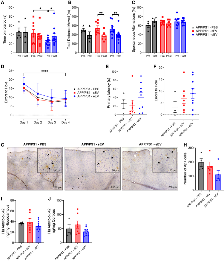
Exercise improves cognitive function in Alzheimer's disease (AD) via mechanism that are not fully clear. Here, we first examined the effect of voluntary exercise training (VET) on energy metabolism and cognitive function in the APP/PS1 transgenic mouse (Tg) model of familial AD. Next, we profiled extracellular vesicles (EVs) and examined whether they may play a role in the protective effects of VET via intranasal administration of EVs, purified from the blood of sedentary (sEV) and/or acutely exercised (eEV) donor wild-type mice into APP/PS1Tg mice. We show that VET reduced resting energy expenditure (REE) and improved cognition in APP/PS1 Tg mice. Administration of eEV, but not sEV, also reduced REE, but had no effect on cognition. Taken together, these data show that exercise is effective intervention to improve symptoms of AD in APP/PS1Tg mice. In addition, eEVs mediate some of these effects, implicating EVs in the treatment of age-related neurodegenerative diseases.

Keywords: Cell biology; Molecular physiology; Neuroscience.


Conflict of interest statement

M.A.F. is a shareholder and scientific advisor for N-Gene Pharmaceuticals. M.A.F. is the founder and shareholder of Celesta Therapeutics.

Figures

None
Graphical abstract
Figure 1
Figure 1
Voluntary exercise training normalizes elevated resting energy expenditure, reduces fat mass, and restores metabolic parameters in male APP/PS1Tg mice to wild-type levels (A) Experimental design. (B) Running wheel data, shown as wheel rotations for a 24-h cycle averaged over three weeks. (C) Total distance over 24 h, averaged over three weeks for both dark and light cycles. (D) Total caloric intake over 48 h during metabolic phenotyping. (E) Body weight. (F) Absolute fat mass. (G) Absolute lean mass; pre-wheels (5 months old) and post-wheels (12 months old). (H and I) (H) Total and (I) resting energy expenditure over same 48 h period, normalized to lean mass. (J) Averaged energy expenditure (hour bins) over same 48-h period, normalized to lean mass. Significance was calculated using two-way/mixed model ANOVA Tukey post hoc, ∗p < 0.05, ∗∗p < 0.01, ∗∗∗p < 0.001, ∗∗∗∗p < 0.0001. Sedentary WT n = 17–21, exercise WT n = 12–13, sedentary APP/PS1 n = 8–12, exercise APP/PS1 n = 9–13. All data are presented as the group mean ± SEM.
Figure 2
Figure 2
Voluntary exercise training enhances long-term memory in male APP/PS1Tg mice, reduces cortical Aβ accumulation and lowers microglial activation, without affecting short-term memory or motor coordination (A) Rotarod results for all four experimental groups. (B) Total distance moved during locomotor activity test (LMA). (C) Percentage of spontaneous alternations during a spontaneous alternation Y-maze test. (D) Errors to the target hole during the Barnes maze learning phase. (E and F) (E) Primary latency and (F) errors to target hole achieved during probe test conducted using a Barnes maze. (G) Representative heat maps from two mice per group during the probe test of the Barnes maze. The location of the target hole is indicated by red circles. (H) Representative images of DAB-stained cortical regions from sedentary and exercise APP/PS1 mice. DAB staining (brown) indicates the presence of amyloid-beta (Aβ) plaques in the cortex (indicated with black arrows), with hematoxylin counterstain (blue) highlighting cell nuclei. (I) Quantification of Aβ plaque burden across the left cerebral hemisphere in sedentary and exercise APP/PS1 mice. Average number of Aβ-positive cells per section determined by two independent blinded scorers. (J) Protein levels of human amyloid-β42 measured using ELISA in the hippocampus of sedentary and exercised APP/PS1 mice, shown as a ratio to total hippocampal tissue loaded. (K) Protein levels of human amyloid-β42 measured using ELISA in the cortex of sedentary and exercised APP/PS1 mice, shown as a ratio to total cortex tissue loaded. (L) Representative western blot images displaying IBA1 protein expression in brain tissue samples from sedentary and exercise WT and APP/PS1 groups. (M) Quantification of IBA1 protein levels normalized to β-actin and expressed relative to the sedentary WT group. Significance was calculated using two-way ANOVA Tukey post hoc (A–E, M) negative binomial regression Tukey post hoc (F), and unpaired t test (I–K). ∗p < 0.05, ∗∗p < 0.01, ∗∗∗p < 0.001, ∗∗∗∗p < 0.0001. Sedentary WT n = 7–21, exercise WT n = 6–13, sedentary APP/PS1 n = 5–12, exercise APP/PS1 n = 4–13. All data are presented as the group mean ± SEM.
Figure 3
Figure 3
Acute treadmill exercise in C57BL/6 mice releases extracellular vesicles expressing tetraspanin proteins consistent with markers of small EVs (A) Size distribution acquired by ZetaView of exercise and sedentary EVs isolated from single mice using SEC (n = 3 per condition). (B) Transmission electron microscopy (TEM) for each fraction isolated from sedentary mice plasma using the qEV10/35 nm column. (C) TEM for each fraction isolated from exercise mice plasma using SEC. (D) Western blot for various antibodies on individual SEC fractions from exercise and sedentary mouse plasma. (E) Western blot for various antibodies on pooled fractions from EVs isolated from exercise and sedentary mouse plasma. EV-rich (1–3) and non-EV (5–7) SEC fractions were pooled and concentrated using Amicon Ultra-15 Centrifugal Filter Units. Three replicates for each of the pooled fractions. (F) Concentration of corticosterone in sedentary and exercise plasma, EVs, and non-EV fractions isolated via SEC, and positive control consisting of plasma from mice restrained for 2 h. ∗∗p < 0.01, ∗∗∗p < 0.001, ∗∗∗∗p < 0.0001. Sedentary plasma n = 8, exercise plasma n = 8, sedentary EVs n = 2, exercise EVs n = 2, sedentary non-EVs n = 2, exercise non-EVs n = 2. All data are presented as the group mean ± SEM.
Figure 4
Figure 4
Exercise-released extracellular vesicles display a distinct miRNA profile compared to sedentary controls, revealing unique miRNA species that may influence processes relevant to cognitive function and Alzheimer’s disease pathology (A) Venn diagram showing the overlap between differentially expressed EV miRNAs between sedentary and exercise. (B) Top 25 differentially regulated miRNAs (>log2 FC 0.5), upregulated (blue), and downregulated (red) in exercise EVs. (C) Heatmap depicting the differential expression of EV miRNAs in exercise EVs vs. sedentary EVs. Rows correspond to individual miRNAs and their relative expression levels. Red indicates expression levels lower than the mean, whereas green indicates expression levels higher than the mean.
Figure 5
Figure 5
Exercise-released extracellular vesicles are enriched in proteins linked to mitochondrial biogenesis and integrin signaling, highlighting potential pathways relevant to Alzheimer’s disease and neuronal EV uptake (A) Top: Venn diagram showing the overlap between proteins identified in mouse EVs from exercise plasma and markers associated with small EVs, and bottom: Venn diagram showing overlap between proteins identified in EVs and exclusion markers. (B) Venn diagram showing the overlap between proteins identified in mouse EVs from exercise plasma, proteins upregulated in human plasma post-exercise and in EVs isolated from human plasma post-exercise. (C) Pathway enrichment analysis performed on the 84 significantly differentially expressed proteins identified in exercise EVs compared with sedentary EVs. Pathways enriched in exercise EVs that may regulate processes involved in metabolism and drive tropism are highlighted in red. Sedentary EVs were considered controls, and exercise EVs were the condition of interest. The size and color of the horizontal bars indicate the scale of enrichment and level of significance (logarithm of the adjusted p value [FDR], orange to red).
Figure 6
Figure 6
Intranasal delivery of exercise-released extracellular vesicles reduces resting and total energy expenditure in APP/PS1Tg mice, recapitulating the metabolic effects of voluntary exercise training (A) Experimental design. (B) Body weight. (C) Absolute fat mass. (D) Absolute lean mass; pre-treatment (4 months old) and post-treatment (12 months old). (E) Total caloric intake over 48 h during metabolic phenotyping. (F) Total distance moved over 48 h during metabolic phenotyping. (G and H) (G) Total and (H) resting energy expenditure over the same 48 h period, normalized to lean mass. (I) Averaged energy expenditure (hour bins) over same 48-h period, normalized to lean mass. Significance was calculated using two-way/mixed model ANOVA Tukey post hoc, ∗p < 0.05, ∗∗p < 0.01, ∗∗∗p < 0.001, ∗∗∗∗p < 0.0001. APP/PS1 PBS, n = 4; APP/PS1 sEV, n = 7; APP/PS1 eEV, n = 10. All data are presented as the group mean ± SEM.
Figure 7
Figure 7
Intranasal delivery of exercise-released extracellular vesicles improves metabolic parameters but does not enhance cognition or reduce Aβ accumulation in APP/PS1Tg mice (A) Total time spent on rotarod. (B) Total distance moved during locomotor activity test (LMA), pre-treatment (4 months old), and post-treatment (12 months old). (C) Percentage of spontaneous alternations during a spontaneous alternation Y-maze test. (D) Errors to the target hole during the Barnes maze learning phase. (E and F) (E) Primary latency and (F) errors to target hole achieved during probe test conducted during the Barnes maze text. (G) Representative images of DAB-stained cortical regions from PBS and EV treated APP/PS1 mice. DAB staining (brown) indicates the presence of Aβ plaques in the cortex (indicated with black arrows), with hematoxylin counterstain (blue) highlighting cell nuclei. (H) Quantification of Aβ plaque burden across the left cerebral hemisphere in PBS and EV treated APP/PS1 mice. (I) Protein levels of human amyloid-β42 measured using ELISA in the hippocampus of APP/PS1 mice treated with PBS and EVs, shown as a ratio to total hippocampal tissue loaded. (J) Protein levels of human amyloid-β42 measured using ELISA in the hippocampus of APP/PS1 mice treated with PBS and EVs, shown as a ratio to total cortex tissue loaded. Significance was calculated using repeated measures two-way ANOVA Tukey post hoc (A–E), negative binomial regression Tukey post hoc (F) and one-way ANOVA Tukey post hoc (H–J) ∗p < 0.05, ∗∗p < 0.01, ∗∗∗∗p < 0.0001. APP/PS1 PBS, n = 4; APP/PS1 sEV, n = 7; APP/PS1 eEV, n = 10. All data are presented as the group mean ± SEM.

Similar articles

References

    1. Nations U . United Nations; 2016. World Population Ageing 2015 Highlights.
    1. Alzheimer's A Alzheimer's disease facts and figures. Alzheimer's Dement. 2015;11:332. - PubMed
    1. Doorduijn A.S., Visser M., van de Rest O., Kester M.I., de Leeuw F.A., Boesveldt S., Fieldhouse J.L.P., van den Heuvel E.G.H.M., Teunissen C.E., Scheltens P., et al. Associations of AD biomarkers and cognitive performance with nutritional status: the NUDAD project. Nutrients. 2019;11:1161. - PMC - PubMed
    1. Puranen T.M., Pietila S.E., Pitkala K.H., Kautiainen H., Raivio M., Eloniemi-Sulkava U., Jyvakorpi S.K., Suominen M. Caregivers’ male gender is associated with poor nutrient intake in AD families (NuAD-trial) J. Nutr. Health Aging. 2014;18:672–676. - PubMed
    1. Tabet N., Mantle D., Walker Z., Orrell M. Higher fat and carbohydrate intake in dementia patients is associated with increased blood glutathione peroxidase activity. Int. Psychogeriatr. 2005;17:91–98. - PubMed