. 2024 Dec 21;28(2):111670.
doi: 10.1016/j.isci.2024.111670. eCollection 2025 Feb 21.

Pathogenic KIAA0586/TALPID3 variants are associated with defects in primary and motile cilia

Affiliations

Pathogenic KIAA0586/TALPID3 variants are associated with defects in primary and motile cilia

Jacqueline E Taudien et al. iScience. .

Abstract

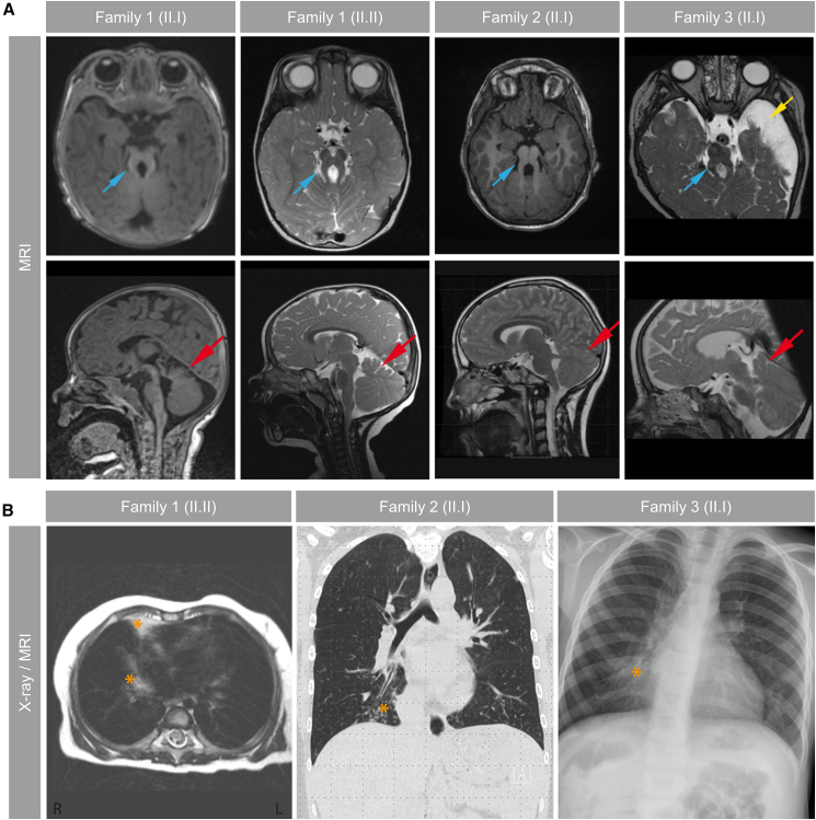
Pathogenic variants in KIAA0586/TALPID3 are associated with the ciliopathy Joubert syndrome (JS). We report individuals with KIAA0586/TALPID3 variants affected by primary and motile cilia defects leading to JS and chronic destructive airway disease. DNA variants were detected in three families by sequencing. In two unrelated families, a deep-intronic variant (KIAA0586/TALPID3:c.3990 + 3186G>A) activated a cryptic exon. We performed histological and functional analyses in native and air-liquid interface (ALI) cultured respiratory cells. Primary cilia lengths were measured in patient-derived fibroblasts. Our data associate KIAA0586/TALPID3 variants with a syndrome combining JS and chronic destructive airway disease, reduced number of motile cilia, disorganized basal body location, and ciliary clearance malfunction. Additionally, patient-derived cell lines showed primary cilia defects. Disease causing KIAA0586/TALPID3 variants, including a deep-intronic sequence variant, were associated with primary and motile cilia defects in JS patients. The combination of JS and respiratory symptoms should be considered indicative for KIAA0586/TALPID3 sequence alterations.

Keywords: Cell biology; Cellular physiology; Human Genetics; Integrative aspects of cell biology.


Conflict of interest statement

The authors declare no competing interests.

Figures

None
Graphical abstract
Figure 1
Figure 1
Segregation analyses of KIAA0586/TALPID3-affected families (A) Illustration of genetic variants identified in KIAA0586/TALPID3 (NM_001329943.3; Chr. 14q23.1; 58,427,385-58,551,297; GRCh38:CM000676.2). Below the chromosome diagram, the KIAA0586/TALPID3 gene is shown as a schematic drawing (light gray bar). The black marking above KIAA0586/TALPID3 represent unaltered exons, while exons with variants are highlighted in green. The blue marking shows the cryptic exon in intron 26. Variants are depicted in red. (B) RT-PCR analyses comparing the patient-derived fibroblast cell line (family 1, index patient [II.I]) with control fibroblasts. The coding region of exon 14 through 20 and exon 22 through 28 were amplified. Induced by the deep-intronic variant KIAA0586/TALPID3:c.(3990 + 3186G>A), the cryptic exon was spliced into the transcript and detected as an additional fragment (marked by a red asterisk). (C) Pedigrees and electropherograms of Sanger sequencing results of three families carrying likely-pathogenic KIAA0586/TALPID3-variants (NM_001329943.3). The following variants cosegregated within families: family 1 c.[2353C>T]; c.[3990 + 3186G>A], family 2 c.[1287delA]; c.[3990 + 3186G>A], and family 3 c.[3785delT]; c.[3785delT]. NTC, non-template control; ref. seq., reference sequence; int, intron; ex, exon; BP, branchpoint; SAS, splice acceptor site; SDS, splice donor site.
Figure 2
Figure 2
KIAA0586/TALPID3-affected individuals show JS-typical findings and destructive airway disease (A) Cranial MRI of all affected individuals showed a molar tooth sign (MTS) (blue arrow). Transversal MRI panels show elongated superior cerebellar peduncles (blue arrow) as well as deepened interpeduncular fossa. Furthermore, patient (II.I) of family 3 additionally was affected by a left media infarct with secondary left-sided brain atrophy (yellow arrow). Midline saggital MRI panels illustrate dysplasia of the cerebellum (red arrow) (cerebellar vermis hypoplasia). (B) MRI of the thorax of patient (II.II) of family 1 revealed middle and lower lobe dystelectasis (orange star). Chest CT of (II.I) of family 2 depicts bronchiectasis as well as mucous plugging (orange star). X-ray of the lungs of patient (II.I) of the family 3 documented chronic middle lobe dystelectasis (orange star).
Figure 3
Figure 3
Transmission electron microscopy (TEM) of KIAA0586/TALPID3-affected respiratory epithelial cells revealed a defect in the generation of multiple motile cilia including reduced numbers of motile cilia, mis-localized basal bodies and bulbous ciliary tips in all four analyzed individuals Respiratory epithelial cells were obtained by a nasal brush biopsy from a healthy control and affected individuals and subsequently analyzed by TEM (family 1 [II.I] TEM analyses after ALI; family 1 [II.II] and family 2 [II.I] TEM analysis directly after nasal brushing, family 3 [II.I] TEM analysis after spheroid cell culture). TEM pictures of the affected show a reduced number of multiple motile cilia, mis-localized basal bodies within the cytoplasm and cilia with bulbous tips. Scale bars represent as noted.
Figure 4
Figure 4
KIAA0586/TALPID3-affected respiratory epithelial cells cultured under ALI-conditions showed reduced number of beating cilia not sufficient to maintain mucociliary clearance in vitro (A) After full differentiation, ALI-cultures were stained with antibodies directed against acetylated alpha-tubulin (acet tub; green) to mark the ciliary axoneme. Nuclei were stained with Hoechst (blue). (B) Left graph: CBF was measured and within normal range for all tested individuals (control: 12 Hz ± 1 Hz; family 1 [II.I]: 11 Hz ± 7 Hz; family 1 [II.II]: 10 Hz ± 3 Hz). Right graph: the calculated active area reflecting ciliary beat movement was markedly reduced in ALI-cultures of the affected individuals (control: 47% ± 13%; family 1 [II.I]: 1% ± 1%; family 1 [II.II]: 1% ± 1%). (C) Statistical analyses of three independent particle tracking experiments per individual revealed reduced speed of transported particles for KIAA0586/TALPID3-affected ALI-cultures (control: 86 μm/s; family 1 [II.I]: 5 μm/s; family 1 [II.II]: 4 μm/s). (D) Particle tracking videos given as z stack projections are exemplary shown per individual. Scale bars in (D) represent 100 μm.
Figure 5
Figure 5
Primary cilia length measurement (A) Images of primary cilia from patient-derived fibroblasts in comparison to two unrelated healthy controls. The axonemes are shown in magenta (detyrosinated tubulin), basal bodies in green (γ-tubulin). Primary cilia of the patient (II.I and II.II) of family 1 showed normal basal bodies and shortend axonemes. (B) The length of primary cilia from patient-derived fibroblasts and from unrelated controls were quantified. In total 750 cilia per cell line were measured in three independent experiments (250 cilia per experiment). The median of the box blots showed 2.6 μm in controls and 1.4 μm and 1.5 μm in the patients. Statistical analysis was performed applying Kruskal-Wallis statistical test. Scale bars: 5 μm, 1 μm for inset; ∗∗∗: p value < 0.0001; ns: not significant.

References

    1. Maria B.L., Hoang K.B., Tusa R.J., Mancuso A.A., Hamed L.M., Quisling R.G., Hove M.T., Fennell E.B., Booth-Jones M., Ringdahl D.M., et al. “Joubert syndrome” revisited: key ocular motor signs with magnetic resonance imaging correlation. J. Child Neurol. 1997;12:423–430. - PubMed
    1. Alby C., Piquand K., Huber C., Megarbané A., Ichkou A., Legendre M., Pelluard F., Encha-Ravazi F., Abi-Tayeh G., Bessières B., et al. Mutations in KIAA0586 Cause Lethal Ciliopathies Ranging from a Hydrolethalus Phenotype to Short-Rib Polydactyly Syndrome. Am. J. Hum. Genet. 2015;97:311–318. - PMC - PubMed
    1. Bachmann-Gagescu R., Dempsey J.C., Phelps I.G., O'Roak B.J., Knutzen D.M., Rue T.C., Ishak G.E., Isabella C.R., Gorden N., Adkins J., et al. Joubert syndrome: a model for untangling recessive disorders with extreme genetic heterogeneity. J. Med. Genet. 2015;52:514–522. - PMC - PubMed
    1. Parisi M., Glass I. In: GeneReviews® [Internet] Adam M.P., Feldman J., Mirzaa G.M., Pagon R.A., Wallace S.E., Amemiya A., editors. University of Washington; Seattle: 2003. Joubert Syndrome; pp. 1993–2024.https://www.ncbi.nlm.nih.gov/books/NBK1325/ - PubMed
    1. Brancati F., Dallapiccola B., Valente E.M. Joubert Syndrome and related disorders. Orphanet J. Rare Dis. 2010;5:20. - PMC - PubMed

LinkOut - more resources