

Intermittent Fasting Reduces Intestinal Inflammation in Dextran Sulfate Sodium-Induced Colitis of Mice
- PMID: 39898122
- PMCID: PMC11787962
- DOI: 10.1002/fsn3.70014
Intermittent Fasting Reduces Intestinal Inflammation in Dextran Sulfate Sodium-Induced Colitis of Mice
Abstract
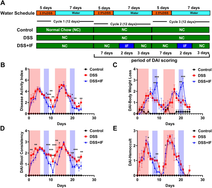
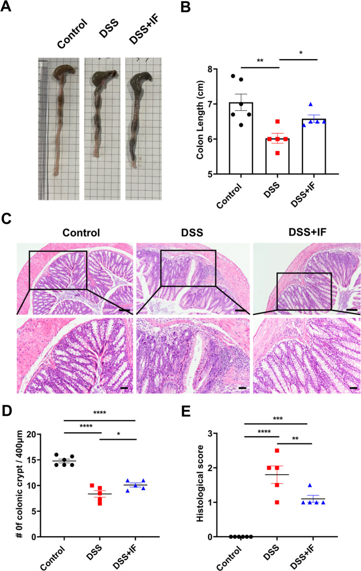
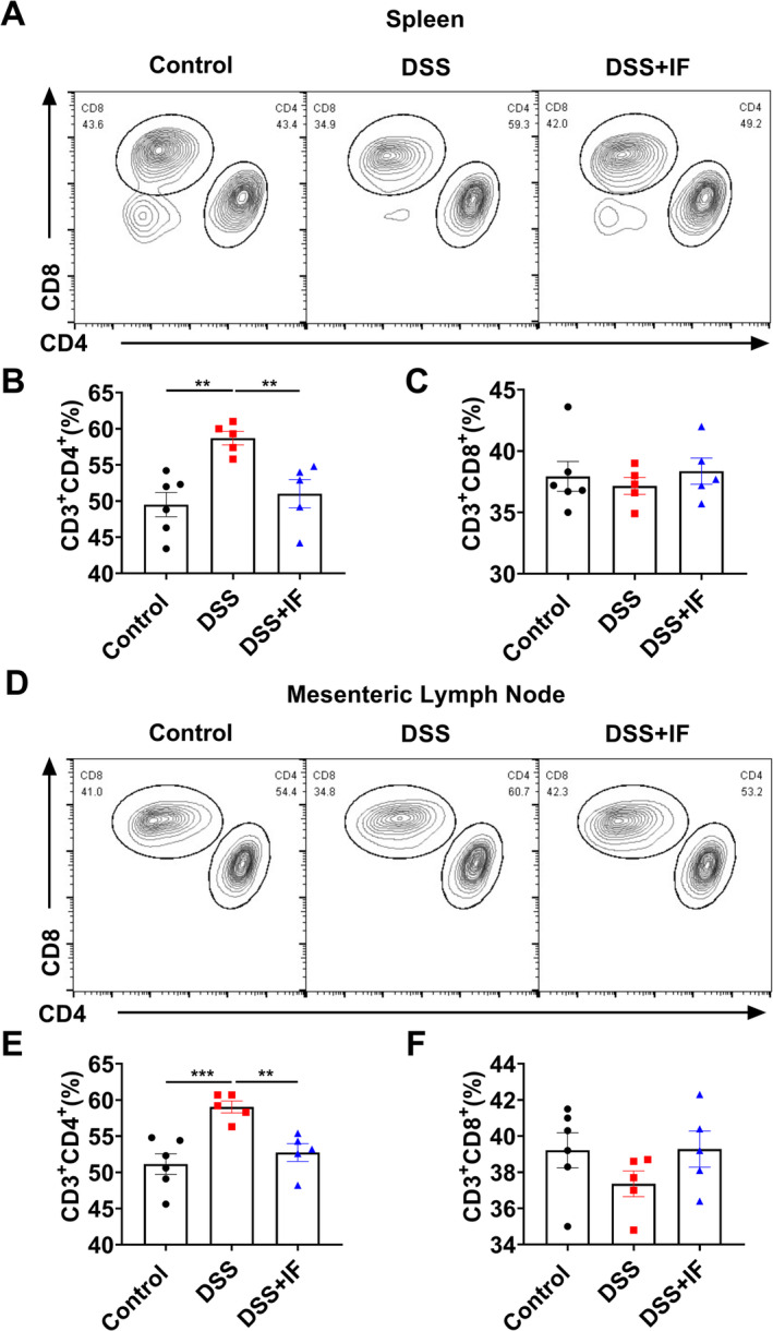
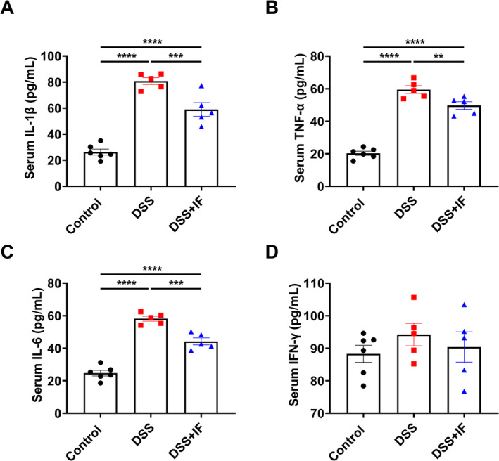
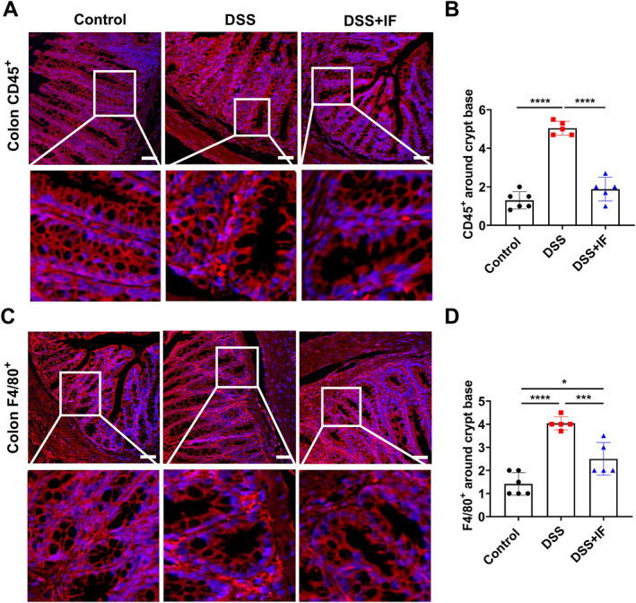
Inflammatory bowel disease (IBD), comprising ulcerative colitis (UC) and Crohn's disease (CD), is a chronic condition impacting both the gastrointestinal tract and the immune system. Intestinal inflammation and epithelial injury are the pathological features of IBD. Recent studies have reported that some strategies of dietary restriction (DR) can regulate immune system, correct the immune disorders, and improve some immune-associated diseases such as IBD. However, as a form of DR, the effect of intermittent fasting (IF) on the IBD remains unknown. In this study, we investigated the therapeutic efficacy of two cycles of IF on the IBD mouse model induced by dextran sulfate sodium (DSS). It was found that two cycles of IF significantly decreased the score of the disease activity index (DAI) and alleviated the IBD-related symptoms. In addition, IF reversed the shortening of colon length mediated by DSS, significantly increased the number of colonic crypts, and decreased the colonic histological score. Furthermore, the proportion of CD4+ T cells in both the spleen and mesenteric lymph node was reduced by IF treatment. The expression of serum pro-inflammatory cytokines IL-1β, TNF-α, and IL-6 was restrained by IF intervention. Moreover, IF administration significantly reduced the number of leukocytes and macrophages infiltrating around the crypt base in the colon. In conclusion, these results demonstrated that IF administration can alleviate the symptoms and pathology of IBD in the DSS-induced IBD mouse model by reducing the intestinal inflammation.
Keywords: inflammation; inflammatory bowel disease; intermittent fasting; intestine.
© 2025 The Author(s). Food Science & Nutrition published by Wiley Periodicals LLC.
Conflict of interest statement
The authors declare no conflicts of interest.
Figures
Similar articles
-
Time-restricted feeding ameliorates dextran sulfate sodium-induced colitis via reducing intestinal inflammation.Front Nutr. 2022 Dec 23;9:1043783. doi: 10.3389/fnut.2022.1043783. eCollection 2022. Front Nutr. 2022. PMID: 36618695 Free PMC article.
-
Investigating intestinal inflammation in DSS-induced model of IBD.J Vis Exp. 2012 Feb 1;(60):3678. doi: 10.3791/3678. J Vis Exp. 2012. PMID: 22331082 Free PMC article.
-
Intermittent administration of a fasting-mimicking diet reduces intestinal inflammation and promotes repair to ameliorate inflammatory bowel disease in mice.J Nutr Biochem. 2021 Oct;96:108785. doi: 10.1016/j.jnutbio.2021.108785. Epub 2021 Jun 1. J Nutr Biochem. 2021. PMID: 34087411
-
2,3,5,4'-Tetrahydroxystilbene-2-O-β-D-glucoside, a major bioactive component from Polygoni multiflori Radix (Heshouwu) suppresses DSS induced acute colitis in BALb/c mice by modulating gut microbiota.Biomed Pharmacother. 2021 May;137:111420. doi: 10.1016/j.biopha.2021.111420. Epub 2021 Feb 23. Biomed Pharmacother. 2021. PMID: 33761623
-
Development, validation and implementation of an in vitro model for the study of metabolic and immune function in normal and inflamed human colonic epithelium.Dan Med J. 2015 Jan;62(1):B4973. Dan Med J. 2015. PMID: 25557335 Review.
References
-
- Bhutani, S. , Klempel M. C., Kroeger C. M., Trepanowski J. F., and Varady K. A.. 2013. “Alternate Day Fasting and Endurance Exercise Combine to Reduce Body Weight and Favorably Alter Plasma Lipids in Obese Humans.” Obesity (Silver Spring) 21, no. 7: 1370–1379. - PubMed
-
- Carter, S. , Clifton P. M., and Keogh J. B.. 2016. “The Effects of Intermittent Compared to Continuous Energy Restriction on Glycaemic Control in Type 2 Diabetes; a Pragmatic Pilot Trial.” Diabetes Research and Clinical Practice 122: 106–112. - PubMed
LinkOut - more resources
Full Text Sources
Research Materials
Miscellaneous
