

Appendiceal Perforation and Abdominal Wall Infection Caused by Invasive Mucormycosis in a Child with Acute Leukemia
- PMID: 39898350
- PMCID: PMC11784252
- DOI: 10.2147/IDR.S504206
Appendiceal Perforation and Abdominal Wall Infection Caused by Invasive Mucormycosis in a Child with Acute Leukemia
Abstract
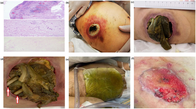
Gastrointestinal mucormycosis is one of the most difficult forms of the disease to diagnose due to its lack of specific clinical features. It is extremely rare to observe gastrointestinal mucormycosis in pediatric acute leukemia patients undergoing chemotherapy. In this report, we describe a case of a child with acute leukemia who developed invasive mucormycosis, leading to appendiceal perforation and abdominal wall infection. Initially, surgical intervention was delayed due to concerns over exacerbating bone marrow suppression, which ultimately resulted in the progression of the intra-abdominal infection. However, after thorough debridement of the abdominal wall infection and treatment with liposomal amphotericin B, the patient gradually recovered. This case highlights the importance of early and complete debridement of abdominal wall infections and intra-abdominal abscesses to prevent the further spread of mucormycosis, shorten the course of the disease, and improve outcomes.
Keywords: abdominal wall infection; acute leukemia; appendiceal perforation; children; invasive mucormycosis.
© 2025 Li et al.
Conflict of interest statement
The authors declare no conflicting interests.
Figures
Similar articles
-
Surgical treatment of appendiceal mucormycosis in an immunocompromised patient: a case report.Surg Case Rep. 2024 Jun 25;10(1):159. doi: 10.1186/s40792-024-01958-y. Surg Case Rep. 2024. PMID: 38916715 Free PMC article.
-
Gastrointestinal Mucormycosis-Induced Massive Lower Gastrointestinal Bleeding, Rectal Perforation, and Pulmonary Embolism: A Long Diagnostic Pathway in a Case Report.Clin Exp Gastroenterol. 2022 Aug 12;15:145-151. doi: 10.2147/CEG.S373728. eCollection 2022. Clin Exp Gastroenterol. 2022. PMID: 35983373 Free PMC article.
-
Post operative abdominal wall mucormycosis infection after laparotomy for bowel perforation.IDCases. 2024 May 24;36:e01998. doi: 10.1016/j.idcr.2024.e01998. eCollection 2024. IDCases. 2024. PMID: 38846026 Free PMC article.
-
Leukemia followed by mixed infection with mucormycosis and aspergillosis: A case report and literature review.Zhong Nan Da Xue Xue Bao Yi Xue Ban. 2023 Jul 28;48(7):1105-1112. doi: 10.11817/j.issn.1672-7347.2023.230039. Zhong Nan Da Xue Xue Bao Yi Xue Ban. 2023. PMID: 37724414 Free PMC article. Review. Chinese, English.
-
[Clinical analysis of 3 cases of mucormycosis in children with acute lymphoblastic leukemia and literature review].Zhonghua Er Ke Za Zhi. 2022 Jan 2;60(1):56-61. doi: 10.3760/cma.j.cn112140-20210711-00571. Zhonghua Er Ke Za Zhi. 2022. PMID: 34986625 Review. Chinese.
References
-
- Pagano L, Offidani M, Fianchi L, et al. Mucormycosis in hematologic patients. Haematologica. 2004;89(2):207–214. - PubMed
Publication types
LinkOut - more resources
Full Text Sources
