

Impact of paternal high energy diets on semen quality and embryo development in cattle
- PMID: 39898513
- PMCID: PMC11850050
- DOI: 10.1530/RAF-24-0082
Impact of paternal high energy diets on semen quality and embryo development in cattle
Abstract
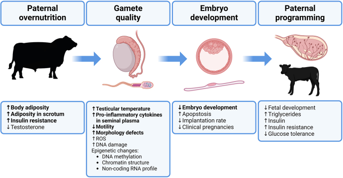
Highly anabolic diets and excessive body fat accumulation have been shown to negatively impact sperm biology in humans and murine biomedical models. Current research indicates that obesity is associated with decreased semen quality and represents a major contributor to male subfertility in humans. Male overnutrition is commonly observed in the beef cattle industry and the use of high energy diets during bull development has been shown to negatively impact semen quality. Most research efforts in bovine reproductive physiology have focused on understanding and optimizing female fertility. This emphasis is even more evident in research investigating the relationship between nutritional interventions and reproductive performance, which has limited the development of nutritional strategies that optimize fertility in bulls. Increasing our understanding of the genetic and environmental factors that influence bull fertility will contribute to future increases in cattle reproductive and productive efficiency. Moreover, exploring the impact of overnutrition in bulls may offer valuable insight and help address diet-induced male subfertility in humans. Herein, we summarize the currently available literature evaluating the impact of highly anabolic diets on male fertility, with an emphasis in the bovine species. Literature summarized in the present review evaluates the impact of overnutrition on sperm biology, early embryonic development, and explores its potential to impact postnatal performance of the offspring.
Conflict of interest statement
The authors declare that there is no conflict of interest that could be perceived as prejudicing the impartiality of the work reported.
Figures


Similar articles
-
Fertility management of bulls to improve beef cattle productivity.Theriogenology. 2016 Jul 1;86(1):397-405. doi: 10.1016/j.theriogenology.2016.04.054. Epub 2016 Apr 22. Theriogenology. 2016. PMID: 27173954 Review.
-
Enhanced early-life nutrition of Holstein bulls increases sperm production potential without decreasing postpubertal semen quality.Theriogenology. 2016 Aug;86(3):687-694.e2. doi: 10.1016/j.theriogenology.2016.02.022. Epub 2016 Mar 4. Theriogenology. 2016. PMID: 27114168
-
Sperm quality in frozen beef and dairy bull semen.Acta Vet Scand. 2018 Jul 4;60(1):41. doi: 10.1186/s13028-018-0396-2. Acta Vet Scand. 2018. PMID: 29973236 Free PMC article.
-
Genetic parameters for various semen production and quality traits and indicators of male and female reproductive performance in Nellore cattle.BMC Genomics. 2023 Mar 27;24(1):150. doi: 10.1186/s12864-023-09216-5. BMC Genomics. 2023. PMID: 36973650 Free PMC article.
-
Review: Genomics of bull fertility.Animal. 2018 Jun;12(s1):s172-s183. doi: 10.1017/S1751731118000599. Epub 2018 Apr 5. Animal. 2018. PMID: 29618393 Free PMC article. Review.
References
LinkOut - more resources
Full Text Sources
