Review
. 2025 Jun;22(6):e202403210.
doi: 10.1002/cbdv.202403210. Epub 2025 Mar 13.

Citrus Flavonoids as Antimicrobials

Affiliations
Review

Citrus Flavonoids as Antimicrobials

Rosaria Ciriminna et al. Chem Biodivers. 2025 Jun.

Abstract

Citrus flavonoids are highly bioactive compounds exerting numerous health benefits including anticancer, antioxidant, antimicrobial, anti-inflammatory, mitoprotective, and neuroprotective activity. Research on their broad-scope bioactivity experienced a renaissance in the early 2000s, and further accelerated after COVID-19, including research on their antimicrobial properties. Summarizing selected research achievements on the antimicrobial activity of the main Citrus flavonoids, this study aims to provide a unified picture on the antimicrobial properties of these valued compounds that will hopefully assist in the development of flavonoid-based antimicrobials, including antibacterial treatments suitable for clinical use minimizing antimicrobial resistance.

Keywords: antimicrobial resistance; citrus flavonoids; diosmin; hesperidin; naringenin; polymethoxyflavones.


Conflict of interest statement

The authors declare no conflicts of interest.

Figures
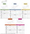
FIGURE 1
FIGURE 1
Flavonoid general structure (top). Flavonoid structures organized by their families. Adapted from [15], Creative Commons Attribution (CC BY) license.
FIGURE 2
FIGURE 2
Documents by year dealing with “citrus flavonoids” indexed by research database Scopus between 1954 and late 2024. Created with data from scopus.com, with kind permission.
FIGURE 3
FIGURE 3
Chemical structures of naringenin (left) and naringin.
FIGURE 4
FIGURE 4
(A) Bacterial levels in the bones of mice from four experimental groups were determined. (B–E) Enzyme‐linked immunosorbent assay was performed to measure the levels of interleukin‐6 (B), IL‐1β (C), C‐reactive protein (D), and tumor necrosis factor‐α (E) in the femurs from each group. Reproduced from [22], with kind permission from John Wiley and Sons, 2024.
FIGURE 5
FIGURE 5
5 Chemical structures of nobiletin (left) and tangeretin.
FIGURE 6
FIGURE 6
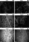
SEM (A–C) and TEM (D–F) photographs showing the effect of nobiletin and tangeretin on morphology of Pseudomonas aeruginosa cells. Reproduced from [25], with kind permission from Elsevier, 2012.
FIGURE 7
FIGURE 7
Antifungal activity of individual pure flavonoid compounds against four phytopathogens at 0.1 mM (A, Sclerotinia sclerotiorum; B, Fusarium oxysporum f. sp. cucumerinum; C, Botrytis cinerea; D, Penicillium digitatum). Reproduced from [27], Creative Commons Attribution 4.0 International License from The Authors, 2021.
FIGURE 8
FIGURE 8
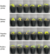
Photos of cucumber seedlings under four treatments after 20 days of cultivation in the greenhouse. Healthy group: cucumber seedlings without any treatment; disease group: cucumber seedlings inoculated with a fungal pathogen Fusarium oxysporum f. sp. cucumerinum and treated with sterilized water; negative group: cucumber seedlings inoculated with the pathogen and treated with empty microcapsules (microcapsules without flavonoid extracts); positive group: cucumber seedlings inoculated with the pathogen and treated with citrus flavonoid microcapsules. Reproduced from [27], Creative Commons Attribution 4.0 International License from The Authors, 2021.
FIGURE 9
FIGURE 9
Chemical structures of hespiridin (left) and hesperetin.
FIGURE 10
FIGURE 10
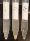
Microbial cells and in culture broth in the presence of hesperetin, hesperidin, and hesperidin glucoside. Reproduced from [32], Creative Commons Attribution (CC BY) license from The Authors, 2022.
FIGURE 11
FIGURE 11
Microbial transformation of hesperidin established in 2023. Reproduced from [33], Creative Commons Attribution (CC BY) license from The Authors, 2023.
FIGURE 12
FIGURE 12
Chemical structure of kaempferol.
FIGURE 13
FIGURE 13
(A) Scheme of the experimental procedures of mice pulmonary infection by Streptococcus pneumoniae. (B) Bacterial burden in the lungs of mice (n = 6 per group) determined by microbiological plating. (C) Pathological observation of lungs in different groups were evaluated by H&E staining. Scale bar = 100 and 200 µm *p < 0.05. [Copyright:  Reproduced from [41] with kind permission from Institut Pasteur, 2022. Published by Elsevier Masson SAS.
FIGURE 14
FIGURE 14
Chemical structures of rutin (left) and its aglycone quercetin (3,3,4,5,7‐pentahydroxiflavone).
FIGURE 15
FIGURE 15
Chemical structures of eriocitrin (left) and eriodictyol.
FIGURE 16
FIGURE 16
Chemical structures of apigenin (left) and apigenin‐7‐O‐glucoside (right).
FIGURE 17
FIGURE 17
Vicenin‐2 on PI3K, AKT, and PTEN signaling in Helicobacter pylori‐infected GES‐1 cells. (A) Western blot analysis of PI3K, AKT, and PTEN expression in vicenin‐2 and/or H. pylori‐infected GES‐1 cells; β‐actin was used as a normal loading control. (B) The quantification of protein was performed by densitometric analysis using ImageJ software. The densitometry data represent means ± SD from three immunoblots and are shown as the comparative density of protein bands normalized to β‐actin. Values not sharing common marking differ significantly at *,# p ≤ 0.05 (Duncan's multiple range test). AKT, protein kinase B; PI3K, phosphatidylinositol 3‐kinase; PTEN, phosphatase and tensin homolog. Reproduced from [49], with kind permission.
FIGURE 18
FIGURE 18
Chemical structures of diosmin.
FIGURE 19
FIGURE 19
Representative CLSM images of Pseudomonas aeruginosa PAO1 biofilms in control conditions (A–D) and after treatment with diosmin (E–H), myricetin (I–L), and neohesperidin (M–P). Bar = 25 µm for A, B, C, E, F, G, I, J, K, M, N, and O; bar = 10 µm for D, H, L, and P. Reproduced from [53], Creative Commons Attribution (CC BY) license from The Authors, 2024.
FIGURE 20
FIGURE 20
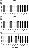
Protective effect of diosmin on survival of embryos after 18, 24, and 48 h post infection (hpi, A, B and C, respectively) with Pseudomonas aeruginosa PAO1 (mean ± SE): ctrl—control group; ictrl—control group with injured tail; B1—107 CFU/mL; B2—106 CFU/mL; M—MIC (0.1 mg/mL); 2M—2 MIC (0.2 mg/mL); str—streptomycin (10 µg/mL). Reproduced from [53], Creative Commons Attribution (CC BY) license from The Authors, 2024.
FIGURE 21
FIGURE 21
Mechanism of action of lemon IntegroPectin films against Gram‐negative bacterial strains: (A) disruption of membrane with consequent leakage of macromolecules in the extracellular environment, (B) targeting of enzymes crucial for cell functioning and vitality, (C) ROS production, (D) inhibition of efflux pumps, (E) acidification of the cytoplasm and protein denaturation, (F) intercalation with the DNA double helix, (G) permeabilization of the bacterial membrane, and (H) inhibition of ATP synthase. Reproduced from [62], Creative Commons Attribution (CC BY) license, from The Authors, 2022.

References

    1. Benavente‐García O., Castillo J., Marin F. R., Ortuño A., and Del Río J. A., “Uses and Properties of Citrus Flavonoids,” Journal of Agricultural and Food Chemistry 45 (1997): 4505–4515, 10.1021/jf970373s. - DOI - PubMed
    1. Search within, “All Fields” Carried Out on scopus.Com, With the Query “Citrus Flavonoids”, up to December 31, 2024. January 31, 2025.
    1. Tripoli E., La Guardia M., Giammanco S., Majo D. D., and Giammanco M., “Citrus Flavonoids: Molecular Structure, Biological Activity and Nutritional Properties: A Review,” Food Chemistry 104 (2007): 466–479, 10.1016/j.foodchem.2006.11.054. - DOI
    1. Alam F., Mohammadin K., Shafique Z., Amjad S. T., and Asad M., “Citrus Flavonoids as Potential Therapeutic Agents: A Review,” Phytotherapy Research 36 (2022): 1417–1441, 10.1002/ptr.7261. - DOI - PubMed
    1. Hooper S. J., Lewis M. A. O., Wilson M. J., and Williams D. W., “Antimicrobial Activity of Citrox Bioflavonoid Preparations Against Oral Microorganisms,” British Dental Journal 210 (2011): E22, 10.1038/sj.bdj.2010.1224. - DOI - PubMed

MeSH terms

LinkOut - more resources