. 2025 Feb 3;82(1):69.
doi: 10.1007/s00018-024-05552-1.

Dnajc5b contributes to male fertility by maintaining the mitochondrial functions and autophagic homeostasis during spermiogenesis

Affiliations

Dnajc5b contributes to male fertility by maintaining the mitochondrial functions and autophagic homeostasis during spermiogenesis

Dake Chen et al. Cell Mol Life Sci. .

Abstract

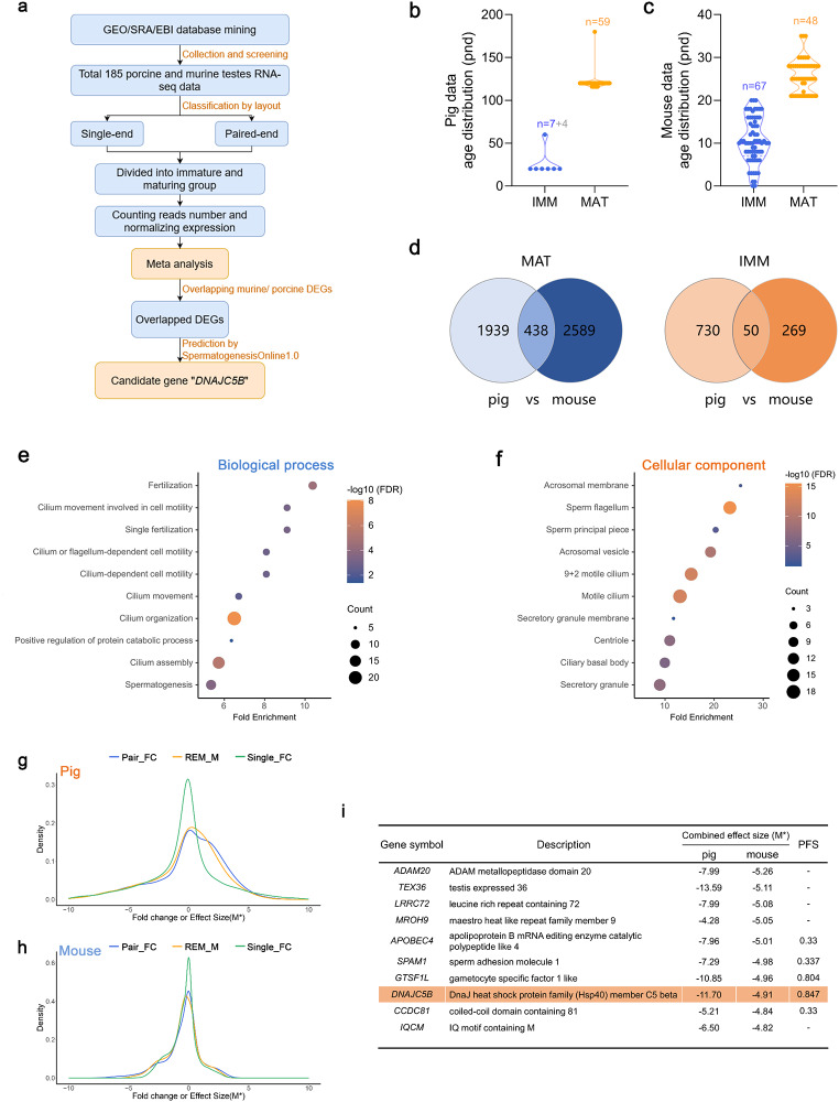
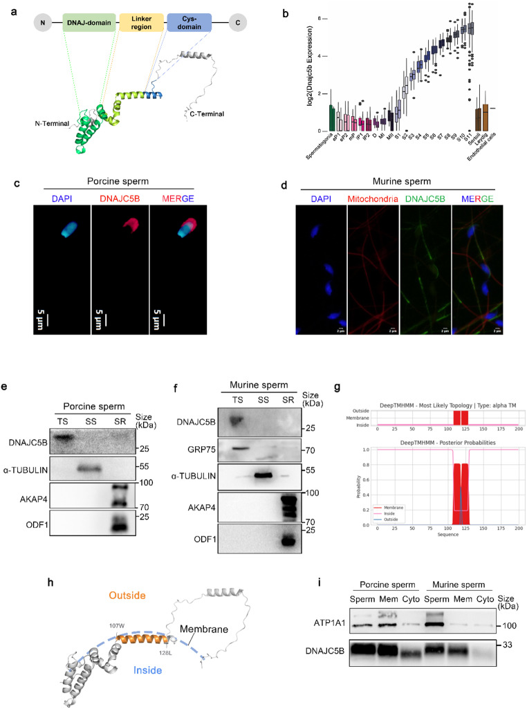
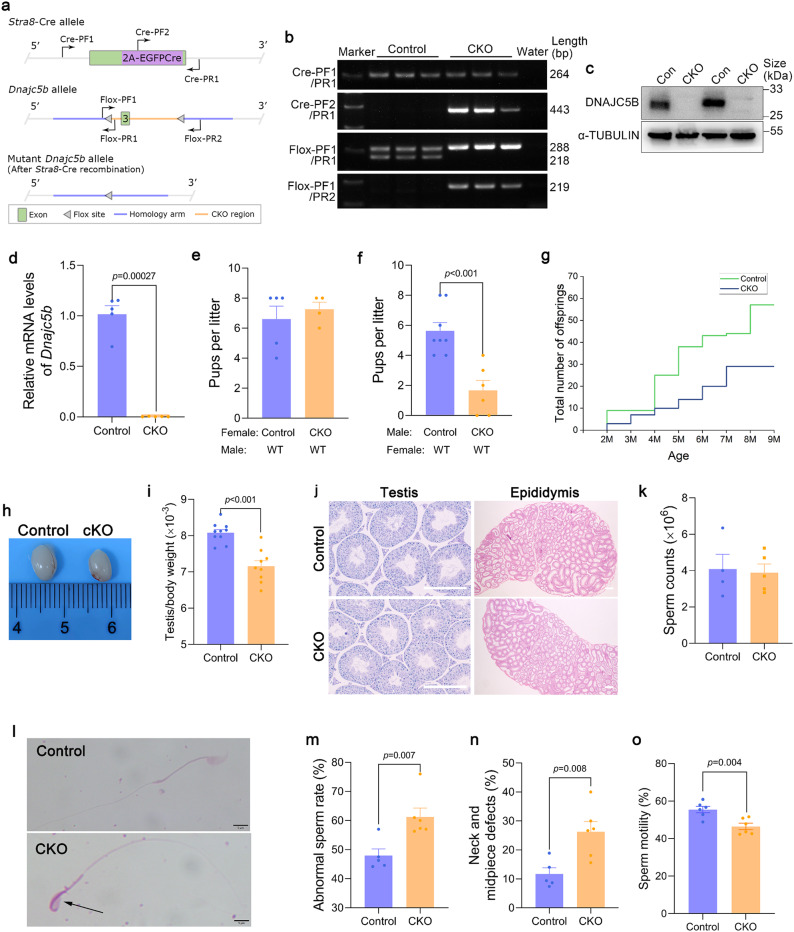
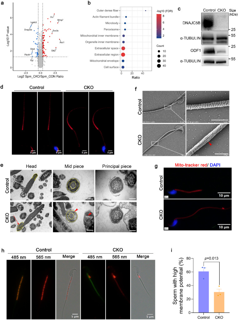
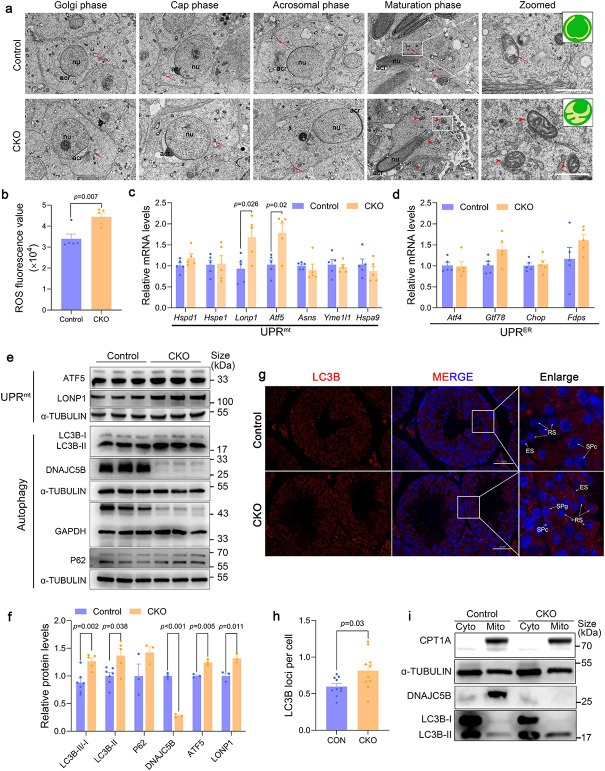
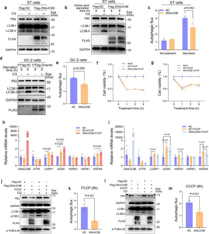
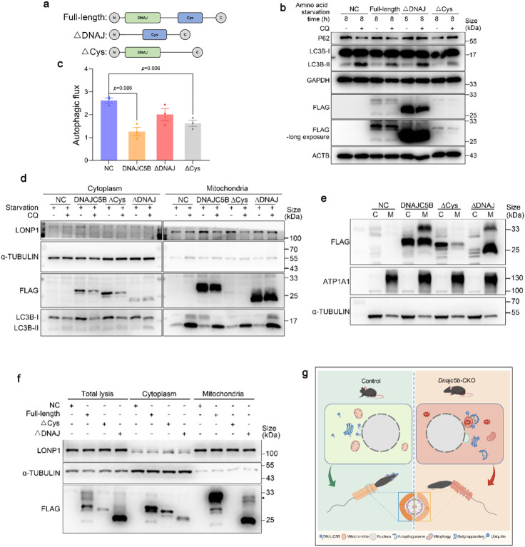
DnaJ heat shock protein family member C5 beta (DNAJC5B), also known as cysteine-string protein beta, exhibits a prominent expression in testicular tissue and plays an important role in acrosomal exocytosis in vitro. Nevertheless, the precise role and underlying mechanism of DNAJC5B in spermatogenesis and male fertility remain poorly understood. The meta-analysis of RNA-sequencing datasets from porcine and murine testes reveals that Dnajc5b could be a pivotal factor in spermatogenesis. This study illustrates that male fertility declines with an increased ratio of abnormal spermatozoa in germ-cell knockout Dnajc5b mice. DNAJC5B has been identified as a mitochondrial protein with high expression in spermatids. The absence of DNAJC5B induces a cascade of mitochondrial damages, including oxidative stress, mitochondrial stress in the testes, and lower mitochondrial membrane potential of spermatozoa. In vivo and in vitro evidence demonstrates that DNAJC5B mitigates excessive cellular autophagy and mitophagy via DNAJ domain under environmental stress conditions, such as starvation or exposure to mitochondrial uncouplers FCCP and CCCP. This study highlights the important role of DNAJC5B in safeguarding male fertility by preserving mitochondrial function and regulating autophagy during spermiogenesis.

Keywords: Dnajc5b; Autophagy; Meta-analysis; Mitochondria; Spermiogenesis.


Conflict of interest statement

Declarations. Ethics approval: Our studies did not include human participants, human data, or human tissue. The animal procedures were approved by the Institutional Animal Care and Use Committee of Huazhong Agricultural University (HZAUMO-2024-0186). Competing interests: The authors have no relevant financial or non-financial interests to disclose.

Figures

Fig. 1
Fig. 1
Meta-analysis shows Dnajc5b is a spermatogenesis-associated gene. a Workflow of data collection, meta-analysis, and candidate genes mining. b The age distribution of porcine samples. n = 11 (IMM, 7 samples have exact age information, and 4 samples are in infancy) and 59 (MAT). pnd: postnatal days. c The age distribution of murine samples. n = 67 (IMM) and 48 (MAT). pnd: postnatal days. d The overlapping DEGs in pig and mouse. There are 438 overlapping DEGs enriched in MAT groups and 50 overlapping DEGs enriched in IMM groups. e, f The biological process (e) and cellular component (f) enrichment analysis of 438 MAT-enriched genes from plot d. g The fold change values and effect sizes distribution of all pig genes. h The fold change values and effect sizes distribution of all mouse genes. i The top 10 MAT-enriched genes ranked by combined effect sizes of 488 murine DEGs. The “PFS” (0–1) was calculated by SpermatogenesisOnline1.0, reflecting the probability of gene function in spermatogenesis
Fig. 2
Fig. 2
DNAJC5B is a membrane-located protein abundantly expressed in spermatids. a The three-dimensional structure of murine DNAJC5B protein predicted by Alphafold2. b Murine Dnajc5b expressions in different cells of murine testes. Dnajc5b expressions were determined by analyzing single-cell RNA-seq data of mature murine testis from the previous study [27]. eP: early pachytene; mP: middle pachytene; lP: late pachytene; D: diplotene; M: metaphase; S: spermatid. c Immunofluorescence assay of DNAJC5B in porcine spermatozoa. The DNAJC5B and nuclei were labeled with DNAJC5B antibody (red) and DAPI (blue), respectively. Scale bars, 5 μm. d Immunofluorescence assay of DNAJC5B in murine cauda spermatozoa. The DNAJC5B, mitochondria and nuclei were labeled with DNAJC5B antibody (green), Mito-Tracker Red CMXRos (red), and DAPI (blue), respectively. Scale bars, 2 μm. e, f Western blot of DNAJC5B in different protein phases from porcine (e) and murine (f) spermatozoa. Proteins in triton-soluble (TS), SDS-soluble (SS) and SDS-resistant (SR) phases were extracted using 1% Triton X-100, 1% SDS and 1× Sample Buffer, respectively. GRP75 is the marker of the triton-soluble phase; αTUBULIN is the marker of the SDS-soluble phase; AKAP4 and ODF1 are the markers of the SDS-resistant phase. g Prediction of transmembrane regions of murine DNAJC5B protein by DeepTMHMM. The region from W107 to L128 of DNAJC5B was predicted as the transmembrane region. h The visualization of murine DNAJC5B protein localization on the membrane by PyMOL software. i Western blot of DNAJC5B in the membrane fraction from murine and porcine spermatozoa. The membrane and cytoplasmic fractions were extracted from murine cauda spermatozoa and porcine spermatozoa. ATP1A1 is the membrane marker of sperm
Fig. 3
Fig. 3
Dnajc5b knockout significantly decreases male mice fertility. a Schematic illustration of the targeting strategy for generating germ cell-conditional Dnajc5b knockout mice. b Genotyping of control and CKO mice by PCR amplification. Primers Cre-PF/PR and Flox-PF/PR were used to amplify the Cre region and the Flox region, respectively. c, d Western blot (c) and qRT-PCR (d) analyses of Dnajc5b expressions in testes of 8-week-old control and CKO mice. n = 2 (WB) and 5 (qRT-PCR). e The mating assays of control and CKO female mice. n = 5 (control) and 4 (CKO). f The mating assays of control and CKO male mice. n = 8 (control) and 6 (CKO). g Total number of offspring during 7-month mating between control or CKO male mice and wild-type female mice. h, i The representative photo (h) and weights (i) of male testes from 8-week-old control and CKO mice. n = 10 (control) and 9 (CKO). j PAS staining of testis and H&E staining of epididymis from 8-week-old control and CKO mice. Scale bars, 200 μm. k The counts of cauda spermatozoa from 8-week-old control and CKO mice. n = 4 (control) and 5 (CKO). l Wright-Giemsa staining of cauda spermatozoa. The arrow indicates abnormal spermatozoa. Scale bars, 5 μm. m, n The abnormal sperm rate (m) and the rate of sperm with abnormal head and neck (n) from control and CKO mice, n = 5 (control) and 6 (CKO). o The motility of spermatozoa in control and CKO mice. n = 6. Data are represented as mean ± SEM of three independent experiments
Fig. 4
Fig. 4
Dnajc5b deficiency impairs the structure and functions of spermatozoa mitochondria. a The differentially expressed proteins (DEPs) in CKO and control spermatozoa. The cutoffs including a fold change greater than 1.30 or less than 0.77 and a p-value less than 0.05 were utilized. b The PANTHER protein class analysis of regulated proteins in CKO spermatozoa. c Western blot of representative proteins (DNAJC5B and outer dense fiber 1) in control and CKO spermatozoa. d Immunofluorescence of DNAJC5B in control and CKO spermatozoa. The signal of DNAJC5B disappeared from the acrosome of CKO spermatozoa. The DNAJC5B and nuclei were labeled with DNAJC5B antibody (red) and DAPI (blue), respectively. Scale bar, 5 μm. e TEM observations of the control and CKO spermatozoa. The arrow indicates abnormal acrosome, and the arrowheads indicate irregular mitochondria. Scale bars, 200 nm. f Scanning electron microscopy observations of the control and CKO spermatozoa. The arrowhead indicates an impaired mitochondrial sheath and the absence of mitochondria in CKO spermatozoa. Scale bars, 2 μm. g Mito-Tracker (red) staining of mitochondria in control and CKO cauda spermatozoa. Scale bars, 10 μm. h, i The representation images of sperm mitochondrial membrane potential (MMP) stained by JC-1 probes (h), and the ratio of high MMP spermatozoa (i) in control and CKO mice. The JC-1 probe can be detected by excitation at 485 nm (green, for the monomeric form) and 565 nm (red, for the aggregated form), reflecting the low or high MMP, respectively. Scale bars, 5 μm. n = 3. Data are represented as mean ± SEM of three independent experiments
Fig. 5
Fig. 5
Loss of Dnajc5b breaks mitochondrial function and activates autophagy in the testis. a TEM observation of the control and CKO testicular spermatids. The red arrows indicate the condensed mitochondria and the red arrowheads indicate the mitochondria with loose matrix. The cartoon models show the condensed mitochondria in control spermatids and the mitochondria with loose matrix in CKO spermatids; the mitochondrial matrix is shown in yellow and the mitochondrial intermembrane space is shown in green. nu: nucleus; acr: acrosome. Scale bars, 1 μm. b The ROS levels of the control and CKO testicular cells. n = 5. c, d The mRNA levels of the genes related to unfolding protein response of mitochondria (UPRmt, c) and endoplasmic reticulum (UPRER, d) in control and CKO testes by qRT-PCR. n = 5. e Western blot of UPRmt proteins (ATF5 and LONP1) and autophagy-related proteins (LC3B and P62) in control and CKO testes. f Quantitative results of protein expression levels in plot e. n = 3. g Immunofluorescence of LC3B in control and CKO testes. The autophagosomes are indicated by LC3B puncta. Scale bars, 50 μm. h The ratio of LC3B puncta per testicular cell. Random areas were selected for counting the number of LC3B puncta. SPg: spermatogonia; SPc: spermatocyte; RS: round spermatid; ES: elongating spermatid; n = 10. i Western blot of LC3B-II and LC3B-I in testicular mitochondria and cytoplasm. CPT1A is the marker of mitochondria. α-TUBULIN is the marker of cytoplasm. Data are represented as mean ± SEM of three independent experiments
Fig. 6
Fig. 6
DNAJC5B negatively regulates the autophagy of cells under starvation or mitochondrial stress. a Western blot of the autophagy-related proteins in ST cells. The autophagy level was consistent in ST cells overexpressing negative control or DNAJC5B. b Western blot of the LC3B protein in ST cells after amino acids starvation. The LC3B-II significantly decreased in DNAJC5B overexpressing ST cells exposed to starvation and 50 µM autophagy inhibitor chloroquine (CQ). c The autophagic flux of ST cells in plot a and plot b. Autophagic flux is reflected by the ratio of (LC3B-II + CQ/GAPDH)/(LC3B-II-CQ/GAPDH). n = 3 (No-treatment) and 4 (starvation). d Western blot of the LC3B protein in GC-2 cells after amino acids starvation. e The autophagic flux of GC-2 cells in plot d. Autophagic flux is reflected by the ratio of (LC3B-II + CQ/GAPDH)/(LC3B-II-CQ/GAPDH). n = 3. f, g Cell viability of ST cells treated with negative or DNAJC5B overexpression vectors, followed by exposure to 10 µM FCCP (a) or 10 µM CCCP (b) for 0, 1, 2, 6 h. n = 6. h, i The relative mRNA expressions of mitochondrial stress-related genes in ST cells treated with DNAJC5B overexpression vectors and 10 µM FCCP (a mitochondrial ROS inducer, h) or 10 µM CCCP (a mitophagy inducer, i) for 6 h. n = 4 (plot h) and 3 (plot i), respectively. j The LC3B protein levels of ST cells exposed to 10 µM FCCP for 6 h. k The autophagic flux in ST cells following treatment of FCCP. The autophagic flux was calculated as the ratio of (LC3B-II + CQ/α-TUBULIN)/(LC3B-II-CQ/α-TUBULIN), corresponding to LC3B-II turnover. n = 3. l The LC3B protein levels in ST cells exposed to 10 µM CCCP for 6 h. m The autophagic flux in ST cells treated with CCCP. The autophagic flux was calculated as the ratio of (LC3B-II + CQ/α-TUBULIN)/(LC3B-II-CQ/α-TUBULIN), corresponding to LC3B-II turnover. n = 3. Data are represented as mean ± SEM of three independent experiments
Fig. 7
Fig. 7
DNAJC5B inhibits cellular autophagy via DNAJ domain. a Construction of porcine DNAJC5B fragments lacking the DNAJ domain or Cys domain. b Western blot of the LC3B in amino acids starved ST cells overexpressing DNAJC5B or its domain-deleted mutants. c The autophagic flux of ST cells in plot b. Autophagic flux is reflected by the ratio of (LC3B-II + CQ/GAPDH)/(LC3B-II-CQ/GAPDH). n = 3. d Western blot of the LC3B in cytoplasm and mitochondria of amino acids starved ST cells overexpressing DNAJC5B or its domain-deleted mutants. e Western blot of the DNAJC5B in cytoplasm and membrane of ST cells overexpressing DNAJC5B or its domain-deleted mutants. Letter “C” is the short form of cytoplasm; Letter “M” is the short form of mitochondria. f Western blot of the DNAJC5B in mitochondria fraction and cytoplasm fraction without mitochondria of ST cells overexpressing DNAJC5B or its domain-deleted mutants. g The schematic diagram of Dnajc5b contributing to spermiogenesis and male fertility by sustaining mitochondrial and autophagic homeostasis. DNAJC5B is localized to the mitochondrial membrane in spermatids and the acrosome in spermatozoa. Male mice deficient in Dnajc5b exhibit subfertility, accompanied by a significantly elevated proportion of abnormal spermatozoa. During spermiogenesis, conditional knockout (CKO) spermatids display mitochondrial dysfunction, characterized by excessive reactive oxygen species (ROS) accumulation and hyperactivated autophagy, thus resulting in damaged spermatozoa

Similar articles

References

    1. Nishimura H, L’Hernault SW (2017) Spermatogenesis. Curr Biol 18:R988–R994. 10.1016/j.cub.2017.07.067 - PubMed
    1. Song H, Zhu L, Li Y, Ma C, Guan K, Xia X, Li F (2015) Exploiting RNA-sequencing data from the porcine testes to identify the key genes involved in spermatogenesis in large White pigs. Gene 2:303–309. 10.1016/j.gene.2015.07.057 - PubMed
    1. Li Y, Li J, Fang C, Shi L, Tan J, Xiong Y, Bin F, Li C (2016) Genome-wide differential expression of genes and small RNAs in testis of two different porcine breeds and at two different ages. Sci Rep 26852. 10.1038/srep26852 - PMC - PubMed
    1. Zhang Y, Xu X, Hu M, Wang X, Cheng H, Zhou R (2021) SPATA33 is an autophagy mediator for cargo selectivity in germline mitophagy. Cell Death Differ 3:1076–1090. 10.1038/s41418-020-00638-2 - PMC - PubMed
    1. Dai J, Zhang T, Guo J, Zhou Q, Gu Y, Zhang J, Hu L, Zong Y, Song J, Zhang S et al (2021) Homozygous pathogenic variants in ACTL9 cause fertilization failure and male infertility in humans and mice. Am J Hum Genet 3:469–481. 10.1016/j.ajhg.2021.02.004 - PMC - PubMed

Substances

LinkOut - more resources