. 2025 Feb 3;149(1):12.
doi: 10.1007/s00401-025-02849-8.

IDH-mutant astrocytomas with primitive neuronal component have a distinct methylation profile and a higher risk of leptomeningeal spread

Affiliations

IDH-mutant astrocytomas with primitive neuronal component have a distinct methylation profile and a higher risk of leptomeningeal spread

Felix Hinz et al. Acta Neuropathol. .

Abstract

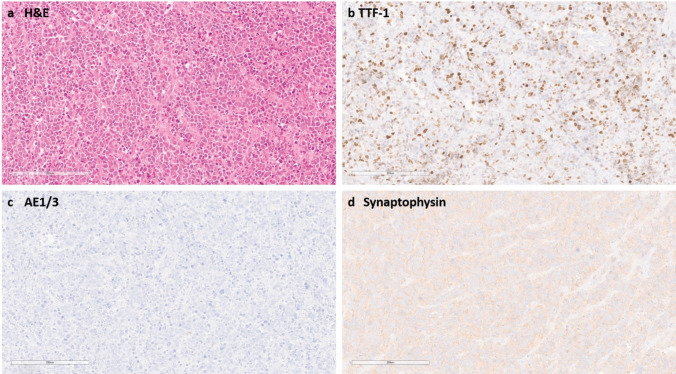
IDH-mutant astrocytomas are diffuse gliomas that are defined by characteristic mutations in IDH1 or IDH2 and do not have complete 1p/19q co-deletion. The established grading criteria include histological features of brisk mitotic activity (grade 3) and necrosis and/or microvascular proliferation (grade 4). In addition, homozygous deletion of the CDKN2A/B locus has recently been implemented as a molecular marker for grade 4 IDH-mutant astrocytomas. Here, we describe a subgroup of high-grade IDH-mutant astrocytomas characterised by a primitive neuronal component based on histology and a distinct DNA methylation profile (n = 51, ASTRO PNC). Misinterpretation as carcinoma metastasis was common, since GFAP expression was absent in the primitive neuronal component, whereas TTF-1 expression was detected in 15/19 cases (79%) based on immunohistochemistry. Apart from mutations in IDH1, TP53, and ATRX, we observed enrichment for alterations in RB1 (n = 19/51, 37%) and MYCN (n = 14/51, 27%). Homozygous CDKN2A/B deletion (n = 1/51, 2%) and CDK4 amplification (n = 3/51, 6%) were relatively rare events. Clinical (n = 31 patients) and survival data (n = 23 patients) indicate a clinical behaviour similar to other CNS WHO grade 4 IDH-mutant astrocytomas, however with an increased risk for leptomeningeal (n = 7) and extra-axial (n = 2) spread. Taken together, ASTRO PNC is defined by a distinct molecular and histological appearance that can mimic metastatic disease and typically follows an aggressive clinical course.

Keywords: MYCN; RB1; Astrocytoma; DNA methylation; IDH-mutant; Primitive neuronal component.


Conflict of interest statement

Declarations. Conflict of interest: MS is scientific advisor and shareholder of Heidelberg Epignostix and Halo Dx, and a scientific advisor of Arima Genomics, and InnoSIGN, and received research funding from Lilly USA, and Illumina USA. DC, SMP, DS, AvD and FS are shareholders of Heidelberg Epignostix. Ethical approval: Collection and analysis of corresponding tissue samples and clinical data was performed in accordance with local ethics regulations (local ethics vote S-318/2022) and in accordance with the ethical standards as laid down in the 1964 Declaration of Helsinki and its later amendments.

Figures

Fig. 1
Fig. 1
a The index case presents itself with a small blue round cell morphology with mitotic figures and occasional cell wrapping. b TTF-1 immunohistochemistry is expressed in the tumour cells. c High-molecular-weight cytokeratins were not expressed. d Synaptophysin is expressed. Scale bar denotes 200 µm
Fig. 2
Fig. 2
Methylation data: in a tSNE analysis with selected reference entities from our database (n = 310), astrocytoma, IDH-mutant, PNC separates clearly from the other entities. PMMRDIA, primary mismatch-repair-deficient IDH-mutant astrocytoma; MB SHH IDH, medulloblastoma, SHH-activated, IDH-mutant; GBM PNC, glioblastoma, IDH-wildtype, with primitive neuronal component. The methylation class astrocytoma, IDH-mutant, high grade consists of 28 grade 4 tumours and 3 grade 3 tumours. The methylation class astrocytoma, IDH-mutant, low grade consists of 2 grade 4 tumours, 13 grade 3 tumours and 17 grade 2 tumours
Fig. 3
Fig. 3
a All cases investigated by histology harbour a primitive neuronal component with small blue round tumour cells. In addition, in this case, cell wrapping (arrow) can be observed. b In most cases, a sharp demarcation of the primitive neuronal component and the astrocytic component can be found. c Presence of tumour growth inside the lumina of two vessels (asterisks). Despite reported extra-axial spread in this tumour entity, this case does not have reported metastases: d some cases present themselves with rosettes (arrows). Scale bar denotes 60 µm in a, b and d. Scale bar denotes 600 µm in c
Fig. 4
Fig. 4
a TTF-1: the primitive neuronal component expresses TTF-1 (using TTF-1 clone EP229). In a subset of cases, the expression is limited to a small number of cells. b IDH1 R132H: most tumour cases strongly express IDH1 R132H. c ATRX: nuclear ATRX expression is lost in most of the cases with blood vessels suited as positive internal controls. d GFAP: GFAP expression is lost in the primitive neuronal component. e Olig2: a subset of the primitive tumour cells expresses Olig2. f AE1/3: the tumours do not express high-molecular-weight cytokeratins stained with AE1 and/or AE3. g Ki-67: the proliferation index is high in the primitive neuronal component, whilst in most cases, the glial component does not show an elevated proliferation index. h p53: p53 is strongly expressed in the primitive tumour cells. i Synaptophysin: synaptophysin is expressed in the primitive tumour cells. Scale bar denotes 300 µm
Fig. 5
Fig. 5
a Oncoprint of 21 samples for which DNA targeted panel sequencing was performed, including genetic mutations, immunohistochemistry (IHC) and copy number variations calculated from the DNA methylation array. b The copy number summary plot (n = 42) reveals a focal deletion at the RB1 locus in addition to several chromosomal gains and losses
Fig. 6
Fig. 6
a Left side: the most common site of origin for primary diagnoses of ASTRO PNC is the frontal lobe followed by the temporal lobe. Right side: for recurrent ASTRO PNC cases, the most common site is the temporal lobe. b The tumours show a predisposition for the male sex. c Age distribution in ASTRO PNC (n = 30, median age = 38). d Kaplan–Meier estimate indicates no significant difference to IDH-mutant astrocytomas WHO grade 4 (p = 0.24). In contrast, there is a significant difference to IDH-mutant astrocytomas WHO grade 2 and 3 (p(A2) = < 0.0001; p(A3) = < 0.0001)

References

    1. Balanis NG, Sheu KM, Esedebe FN, Patel SJ, Smith BA, Park JW et al (2019) Pan-cancer convergence to a small-cell neuroendocrine phenotype that shares susceptibilities with hematological malignancies. Cancer Cell 36(17–34):e17. 10.1016/j.ccell.2019.06.005 - PMC - PubMed
    1. Banan R, Stichel D, Bleck A, Hong B, Lehmann U, Suwala A et al (2020) Infratentorial IDH-mutant astrocytoma is a distinct subtype. Acta Neuropathol 140:569–581. 10.1007/s00401-020-02194-y - PubMed
    1. Bingle CD (1997) Thyroid transcription factor-1. Int J Biochem Cell Biol 29:1471–1473. 10.1016/s1357-2725(97)00007-1 - PubMed
    1. Brat DJ, Aldape K, Colman H, Figrarella-Branger D, Fuller GN, Giannini C et al (2020) cIMPACT-NOW update 5: recommended grading criteria and terminologies for IDH-mutant astrocytomas. Acta Neuropathol 139:603–608. 10.1007/s00401-020-02127-9 - PMC - PubMed
    1. Capper D, Jones DTW, Sill M, Hovestadt V, Schrimpf D, Sturm D et al (2018) DNA methylation-based classification of central nervous system tumours. Nature 555:469–474. 10.1038/nature26000 - PMC - PubMed

Publication types

Substances