. 2025 Feb 3;41(1):39.
doi: 10.1007/s10565-025-09991-9.

UHRF1 promotes calcium oxalate-induced renal fibrosis by renal lipid deposition via bridging AMPK dephosphorylation

Affiliations

UHRF1 promotes calcium oxalate-induced renal fibrosis by renal lipid deposition via bridging AMPK dephosphorylation

Yushi Sun et al. Cell Biol Toxicol. .

Abstract

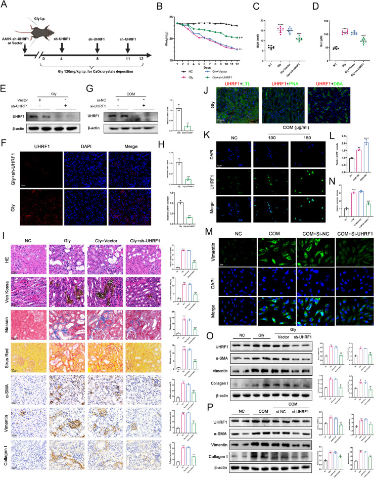
Background: Nephrolithiasis, a common urinary system disorder, exhibits high morbidity and recurrence rates, correlating with renal dysfunction and the increased risk of chronic kidney disease. Nonetheless, the precise role of disrupted cellular metabolism in renal injury induced by calcium oxalate (CaOx) crystal deposition is unclear. The purpose of this study is to investigate the involvement of the ubiquitin-like protein containing PHD and RING finger structural domain 1 (UHRF1) in CaOx-induced renal fibrosis and its impacts on cellular lipid metabolism.

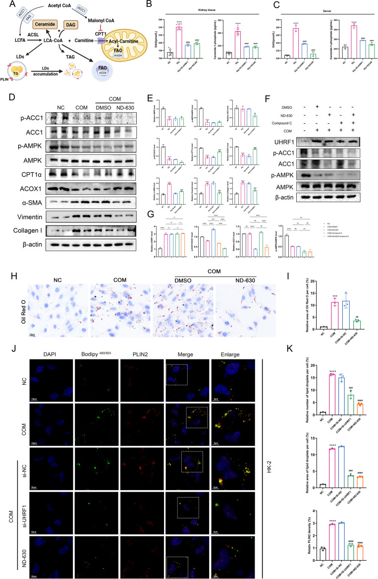
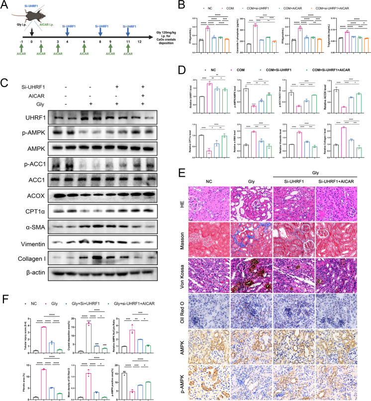
Methods: Various approaches, including snRNA-seq, transcriptome RNA-seq, immunohistochemistry, and western blot analyses, were employed to assess UHRF1 expression in kidneys of nephrolithiasis patients, hyperoxaluric mice, and CaOx-induced renal tubular epithelial cells. Subsequently, knockdown of UHRF1 in mice and cells corroborated its effect of UHRF1 on fibrosis, ectopic lipid deposition (ELD) and fatty acid oxidation (FAO). Rescue experiments using AICAR, ND-630 and Compound-C were performed in UHRF1-knockdown cells to explore the involvement of the AMPK pathway. Then we confirmed the bridging molecule and its regulatory pathway in vitro. Experimental results were finally confirmed using AICAR and chemically modified si-UHRF1 in vivo of hyperoxaluria mice model.

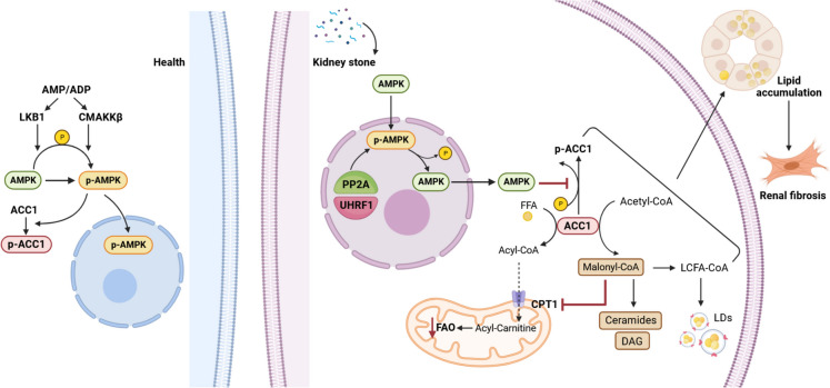
Results: Mechanistically, UHRF1 was found to hinder the activation of the AMPK/ACC1 pathway during CaOx-induced renal fibrosis, which was mitigated by employing AICAR, an AMPK agonist. As a nuclear protein, UHRF1 facilitates nuclear translocation of AMPK and act as a molecular link targeting the protein phosphatase PP2A to dephosphorylate AMPK and inhibit its activity.

Conclusion: This study revealed that UHRF1 promotes CaOx -induced renal fibrosis by enhancing lipid accumulation and suppressing FAO via inhibiting the AMPK pathway. These findings underscore the feasible therapeutic implications of targeting UHRF1 to prevent renal fibrosis due to stones.

Keywords: AMPK; Fatty acid oxidation; Lipid synthesis; Renal fibrosis; UHRF1.


Conflict of interest statement

Declarations. Ethics approval and consent to participate: The Ethics Committee of the Renmin Hospital of Wuhan University approved the clinical specimen collection (approval number: WDRY2021-KS047). The Experimental Animal Welfare and Ethics Committee of Wuhan University People's Hospital (approval number: WDRM20200604) permitted all animal experiments. Consent for publication: Not applicable. Conflicts of interest: The authors declare no competing interests.

Figures

Fig. 1
Fig. 1
(A) RNA transcriptome heatmap showing DNA methylation–related genes between hyperoxaluric mice and healthy controls. High-expression and low-expression genes are respectively shown in red and blue. (B) Volcano plot of DEGs in the Gly group when compared with those in the control. (C) mRNA expression levels of DEGs in panel A. (D) Dnmt3b, Mbd2, Dnmt1, Pcna, Ctcf, and Mecp2 protein levels. (E) snRNA-Seq results showing that UHRF1 expression was upregulated in the nuclei of proximal RTECs in CKD. (F-G) Immunohistochemistry and (H) RT-qPCR showing that UHRF1 expression in the renal tissues of patients with renal calculi was higher than that of healthy individuals. Scale bar = 20 μm. (I-J) Immunofluorescence detection of the renal tissue fibrosis marker molecules α-SMA, Vimentin, and Collagen I in patients with renal calculi, and quantification of the percentage of positive area. Scale bar = 20 μm. (K) Double immunofluorescent staining was performed on the kidney tissues of mice in control and Gly groups. (L) Correlation analysis of UHRF1 and ECM molecules in terms of expression levels. *P < 0.05, ****P < 0.0001 compared with the control group
Fig. 2
Fig. 2
(A) Mouse modeling diagram. (B) Effect of sh-UHRF1 on body weight (n = 6). (C-D) SCr and BUN levels of mice in each group (n = 6). (EF, H) Western blot and immunofluorescence (IF) to detect the knockdown efficiency of sh-UHRF1 in vivo. Scale bar = 20 μm. (I) Percentage of pathological staining and immunochemistry positive area was calculated. Scale bar = 20 μm. (J) Colocalization results of UHRF1 with markers in each renal tubular segment (LTL, PNA and DBA). Scale bar = 20 μm. (K-L) 24 h after COM treatment at different concentrations. IF staining showing the expression level of UHRF1 in HK-2 cells. (G) Western blot to detect the knockdown efficiency of si-UHRF1 in HK-2 cells. (M-P) IF and western blot detected the expression levels of fibrotic molecules in HK-2 cells (MO) and kidney tissues in the Gly group after 150 μg/mL COM treatment. Scale bar = 20 μm. **P < 0.01, ***P < 0.001, ****P < 0.0001 vs control group; #P < 0.05, ##P < 0.01, ###P < 0.001, ####P < 0.0001 vs Gly group
Fig. 3
Fig. 3
(A-B) KEGG and GO enrichment analysis of RNA-seq data. (C) Nile red staining showing that the site of lipid deposition in the Gly group was co-localized with LTL in the proximal RTECs. (D-F) Representative images of lipid accumulation in the kidney of hyperoxaluric mice. (G-H) Representative images of Oil Red O staining in HK-2 cells. (I) OCR of NC, COM and COM + si-UHRF1 HK-2 cells were measured with a Seahorse XFe96 analyser. Basal respiration (J), ATP production (K), maximal respiration (L), and FAO level (M) of 3 groups. n = 6 biologically independent samples. Scale bar = 20 μm. ****P < 0.001 compared with the control group; ####P < 0.0001 compared with the Gly group
Fig. 4
Fig. 4
(A) The GO-BP term "negative regulation of lipid metabolic processes" enriched in Gly group. (B-C) Relative protein expression of key molecules of the UHRF1 and AMPK pathways of lipid metabolism in renal tissue lysates of control, Gly, and sh-UHRF1-treated mice. (D-E) BODIPY493/503 staining pictures of LDs in HK-2 cells. Scale bar = 20 μm. (F-I) Renal cortex samples from each group were examined by ORO staining. (F) HK-2 cells were treated with Compound C and si-UHRF1 for rescue experiments. (I-J) Representative TEM images showed the sizes and numbers of LDs. scale bar for TEM = 2 μm and 1 μm. (G-H) Scale bar for ORO staining = 20 μm. (K) Renal and serum TG and FFAs levels were determined in the indicated groups. ***P < 0.001, ****P < 0.0001 vs. control; ##P < 0.01, ###P < 0.001, ####P < 0.0001 vs. Gly group
Fig. 5
Fig. 5
(A) Graphical representation of intracellular fatty acid synthesis and LDs formation. (B-C) DAG and C-1-P contents in the kidney tissue and serum in the indicated groups. (D-E) Relative protein levels of the AMPK/ACC1 pathway and CPT1α, ACOX1, Col-I, Vimentin, and α-SMA in the renal tissues.(F-G) Recovery experiments using ND-630 and Compound-C. (H-I) ORO detection of the effect of ND-630 on lipid deposition in COM-stimulated HK-2 cells. (J-K) Laser confocal images of BODIPY493/503 and PLIN2 were obtained under each set of conditioned treatments, and LDs and PLIN2 fluorescence was quantified. **P < 0.01, ***P < 0.001, ****P < 0.0001 compared with the control group; ##P < 0.01, ###P < 0.001, ####P < 0.0001 compared with the Gly group
Fig. 6
Fig. 6
(A) Whole-cell extracts of COM-stimulated HK-2 cells were immunoprecipitated using AMPK antibody and immunoblotted using PP2A and UHRF1 antibodies. (B) Si-UHRF1 reduced the extent of PP2A interaction with AMPK. (C-D) In the presence or absence of Si-UHRF1 or Si-PPP2CA conditions, UHRF1, PP2A, and AMPK/ACC1 pathways were detected using western blot and quantified using densitometry. (EF) UHRF1 expression levels in the control, COM, and Si-UHRF1 groups captured using a laser confocal microscope. (EF) Representative images of UHRF1 and AMPK in the indicated groups were captured using a laser confocal microscope. Quantified by densitometry of the relative ratio of the red fluorescence intensity of AMPK in the nucleus to that of the cytoplasm and the relative fluorescence intensity of UHRF1. ***P < 0.001, ****P < 0.0001 compared with the control group; ##P < 0.01, ####P < 0.0001 compared with the Gly group
Fig. 7
Fig. 7
(A) Si-UHRF1 combined with AICAR administration strategy in glyoxylate-induced renal calculi mouse. (B) COM-stimulated HK-2 cells were treated with si-UHRF1, AICAR, and the two in combination, and TG and FFAs contents were measured in each group. (C-D) Western blot results of UHRF1, AMPK/ACC1 pathway, FAO key molecules, and fibrosis markers in mouse kidney tissues. (EF) HE, Von-Kossa, Masson, and ORO staining was used to evaluate renal tubular injury, CaOx deposition, interstitial fibrosis, and lipid accumulation, respectively. Immunohistochemistry was used to detect AMPK and p-AMPK expression. *P < 0.05, **P < 0.01, ***P < 0.001, ****P < 0.0001
Fig. 8
Fig. 8
Under normal conditions (left side), AMPK is activated by LKB1, CAMKKβ, or AMP/ADP, which activates the AMPK pathway. In the presence of kidney stones (right side), CaOx-stimulated elevated expression of UHRF1 in RTECs, which, as a nuclear protein, bridged AMPK dephosphorylation by PP2A and increased nuclear translocation and nuclear retention of AMPK. Inhibition of the AMPK/ACC1 pathway promotes the synthesis of Mal-CoA and FFAs in the cytoplasm, leading to the accumulation of triglycerides and LDs; however, excessive accumulation of Mal-CoA is converted to lipotoxic substances such as ceramides and DAG; and finally, Mal-CoA acts as an endogenous inhibitor of CPT1α and reduces FAO. Disturbances in these lipid metabolic processes promote lipid accumulation in RTECs and renal interstitial fibrosis

Similar articles

References

    1. Alexander RT, Fuster DG, Dimke H. Mechanisms underlying calcium nephrolithiasis. Annu Rev Physiol. 2022;84:559–83. 10.1146/annurev-physiol-052521-121822. - PubMed
    1. Bandet CL, Tan-Chen S, Bourron O, Le Stunff H, Hajduch E. Sphingolipid metabolism: New insight into ceramide-induced lipotoxicity in muscle cells. Int J Mol Sci. 2019;20(3). 10.3390/ijms20030479. - PMC - PubMed
    1. Brownsey RW, Zhande R, Boone AN. Isoforms of acetyl-CoA carboxylase: structures, regulatory properties and metabolic functions. Biochem Soc Trans. 1997;25(4):1232–8. - PubMed
    1. Chen W, Liu WR, Hou JB, Ding JR, Peng ZJ, Gao SY, Dong X, Ma JH, Lin QS, Lu JR, Guo ZY. Metabolomic analysis reveals a protective effect of Fu-Fang-Jin-Qian-Chao herbal granules on oxalate-induced kidney injury. Bioscience Rep. 2019;39(2):BSR20181833. - PMC - PubMed
    1. Cheng D, Wang Y, Li Z, Xiong H, Sun W, Xi S, Zhou S, Liu Y, Ni C. Liposomal UHRF1 siRNA shows lung fibrosis treatment potential through regulation of fibroblast activation. JCI insight. 2022;7(22). 10.1172/jci.insight.162831. - PMC - PubMed

MeSH terms

LinkOut - more resources