Review
. 2025 Feb 3;28(2):15.
doi: 10.1007/s10456-024-09953-5.

From bench to bedside: murine models of inherited and sporadic brain arteriovenous malformations

Affiliations
Review

From bench to bedside: murine models of inherited and sporadic brain arteriovenous malformations

Ashely R Ricciardelli et al. Angiogenesis. .

Abstract

Brain arteriovenous malformations are abnormal vascular structures in which an artery shunts high pressure blood directly to a vein without an intervening capillary bed. These lesions become highly remodeled over time and are prone to rupture. Historically, brain arteriovenous malformations have been challenging to treat, using primarily surgical approaches. Over the past few decades, the genetic causes of these malformations have been uncovered. These can be divided into (1) familial forms, such as loss of function mutations in TGF-β (BMP9/10) components in hereditary hemorrhagic telangiectasia, or (2) sporadic forms, resulting from somatic gain of function mutations in genes involved in the RAS-MAPK signaling pathway. Leveraging these genetic discoveries, preclinical mouse models have been developed to uncover the mechanisms underlying abnormal vessel formation, and thus revealing potential therapeutic targets. Impressively, initial preclinical studies suggest that pharmacological treatments disrupting these aberrant pathways may ameliorate the abnormal pathologic vessel remodeling and inflammatory and hemorrhagic nature of these high-flow vascular anomalies. Intriguingly, these studies also suggest uncontrolled angiogenic signaling may be a major driver in bAVM pathogenesis. This comprehensive review describes the genetics underlying both inherited and sporadic bAVM and details the state of the field regarding murine models of bAVM, highlighting emerging therapeutic targets that may transform our approach to treating these devastating lesions.

Keywords: AVM; BMP; Cerebrovascular; Intracranial hemorrhage; Notch; RAS/MAPK; TGF-β.


Conflict of interest statement

Declarations. Competing interest: The authors declare no competing interests.

Figures

Fig. 1
Fig. 1
Arteriovenous Malformations. A Schematic of a normal arteriovenous network, where arteries and veins only communicate at the intervening capillary bed, where diffusion of oxygen, nutrients, and hormones to surrounding tissues occurs (such as the brain parenchyma). B An arteriovenous malformation [AVM] is the result of a direct connection between a high-flow arterial vessel and a low resistance capacitance venous vessel, with a demarcated nidus (or tangle), and extensive dilation of the draining veins, and tortuous remodeling of both the feeding arteries and draining veins. These lesions can lead to decreased flow through the surrounding capillary vessels (the “steal” phenomenon), and results in local hypoxia surrounding the AVM due to decreased capillary perfusion of adjacent regions. EC = endothelial cell; RBC = red blood cell
Fig. 2
Fig. 2
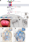
The molecular and genetic etiology of Hereditary Hemorrhagic Telangiectasia (HHT). A HHT is an autosomal dominant inherited syndrome that results from impaired transforming growth factor-β (TGF-β) signaling. Mutations have been observed in genes encoding multiple proteins of this pathway. Loss of function variants in GDF2, which encodes the ligand bone morphogenetic protein 9 (BMP9) lead to HHT type 5 (HHT5) and account for < 1% of all HHT cases, while loss of function mutations in the genes ACVLR1 and ENG, which encode the TGF-β co-receptors activin receptor-like kinase 1 (ALK1) (HHT2) and endoglin (ENG) (HHT1), together account for approximately 90% of all cases of HHT. Lesions in MADH4, which encodes the transcription factor SMAD4, underly juvenile polyposis and HHT (JP-HHT) and are found in 1–2% of all HHT cases. A single study recently reported HHT like symptoms in patients harboring suspected pathogenic, damaging variants in the class 2 double stranded RNA-specific endoribonuclease DROSHA, which executes the initiation step of microRNA processing within the nucleus, and may specifically regulate a class of BMP-target microRNAs. B In HHT patients, small microscopic AVMs, known as telangiectasias (indicated by the black arrows), present on mucosal membranes, such as the finger nails or tongue. C Lateral view of a digital subtractive angiogram of a 3-year-old patient with an HHT mutation, following intracerebral artery (ICA) injection. Note the large left parietal AVM and small poster temporal AVM. Red caret = feeding arteries; blue caret = draining veins; asterisk = AVM. D Coronal (or anteroposterior) view. E Dorsal view of a P7 Alk1flox/flox mouse following blue latex perfusion through the arterial circulation, shows normal formation of the arterial cerebrovasculature. F Same view of an Alk1 induced endothelial cell knockout (Alk1iECKO) littermate, a model of HHT2, with obvious perfusion of the venous vasculature in the brain, indicative of arteriovenous shunts. The boxed in, magnified view shows a perfused artery (red caret) and a perfused vein (blue caret), which is not observed in control animals. The white asterisk denotes the vascular anomaly. BA = basilar artery; C.o.W. = Circle of Willis; br. st. = brain stem; MCA = midcerebral artery; ob = olfactory bulb
Fig. 3
Fig. 3
The Notch signaling pathway. The Notch receptor is transcribed and translated and the polypeptide exported from the cell, where serine and threonine residues within the epidermal growth factor (EGF) repeats of the extracellular domain are O-fucosylated and/or O-glucosylated by the enzymes Pofut and Poglut, respectively, two essential modifications for generating a fully functional receptor. The receptor is further modified by the glycosyltransferase Fringe, as well as xylotransferases, and this “sugar code” on the EGF repeats of the extracellular domain of Notch alters the affinity of the receptor for different ligands. While in the golgi, the polypeptide is proteolytically cleaved by Furin (S1). The cleaved heterodimer is assembled and held together via non-covalent interactions and trafficked to the cell surface. Binding to one of the Delta, Serrate, or Jagged (DSL) ligands on an adjacent cell leads to receptor activation via juxtacrine signaling. Briefly, ligand tension and subsequent endocytosis generate the force necessary to induce a conformational change in the bound receptor that exposes site 2 (S2) for cleavage by ADAM metalloproteases. This cleavage generates the membrane anchored Notch extracellular truncation (NEXT) fragment, a substrate for the γ-secretase complex. γ-secretase cleavage at site 3 (S3) and (S4) results in release of the Nβ peptide and the Notch intracellular domain (NICD). In the absence of Notch signaling, RBPjκ (CBF1/CSL/Su(H)/Lag-1) associates with corepressor proteins and histone deacetylases to repress transcription of target genes. Following ligand activation and receptor cleavage, the NICD translocates to the nucleus and binds to RBPjκ to displace the repressors and recruit the coactivator Mastermind (MAML), which in turn recruits additional coactivators to stimulate transcription. Adapted from Kopan and Ilagan, 2009 [112]
Fig. 4
Fig. 4
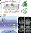
Mutant KRAS and Brain Arteriovenous Malformations. A The sequence identity and conservation of key functional domains across NRAS, HRAS, KRAS4A, and KRAS4B is shown. Somatic mutations described in the literature within KRAS are indicated at the bottom. Similar to known oncogenic mutations in RAS, they occur at codons 12, 13, and 61 and lead to constitutive GTPase activity. B Sequence similarity between the amino terminus of the four RAS isoforms, with protein domains highlighted. Mutations within and around loops 1, 2, and 4 (in Gly12, Gly13, and Glu61) affect nucleotide binding, resulting in enhanced GTP binding by RAS. C Overview of the 3D structure of RAS with the allosteric lobe shown in grey, the effector lobe highlighted in green, and GDP shown in red. D A structural view shows how these residues (Gly12, Gly13, Glu61) within the switch I domain (effector lobe) interact with GDP and GTP. Mutation within these residues prevents GTP hydrolysis, locking RAS in a constitutively active state. E Diagram of a receptor tyrosine kinase pathway, specifically VEGF-VEGFR2 signaling, that acts upstream of KRAS in the endothelium, and the molecular consequence of constitutive KRAS activity favors RAS-MAPK signaling in these cells. F–K Computed micro tomography (micro-CT) imaging of the cerebrovasculature of CNS-endothelial specific KrasG12D mutant mice at 2 months of age following perfusion with a contrast agent reveals striking arteriovenous connections, or arteriovenous shunts, with a dilated feeding artery and draining veins (blue caret) as well as reduced small vessels in the cortex (asterisk). From Suarez et al. 2024 [163]. F, G Dorsal view: H, I sagittal view; J, K coronal view. Panels A and B are adapted from Prior IA et al. 2012 [164]

Similar articles

Cited by

References

    1. Dalton A, Dobson G, Prasad M, Mukerji N (2018) De novo intracerebral arteriovenous malformations and a review of the theories of their formation. Br J Neurosurg 32(3):305–311. 10.1080/02688697.2018.1478060 - PubMed
    1. Morris Z, Whiteley WN, Longstreth WT Jr, Weber F, Lee YC, Tsushima Y, Alphs H, Ladd SC, Warlow C, Wardlaw JM, Al-Shahi Salman R (2009) Incidental findings on brain magnetic resonance imaging: systematic review and meta-analysis. BMJ 339:b3016. 10.1136/bmj.b3016 - PMC - PubMed
    1. Gabriel RA, Kim H, Sidney S, McCulloch CE, Singh V, Johnston SC, Ko NU, Achrol AS, Zaroff JG, Young WL (2010) Ten-year detection rate of brain arteriovenous malformations in a large, multiethnic, defined population. Stroke 41(1):21–26. 10.1161/STROKEAHA.109.566018 - PMC - PubMed
    1. Meyer-Heim AD, Boltshauser E (2003) Spontaneous intracranial haemorrhage in children: aetiology, presentation and outcome. Brain Dev 25(6):416–421. 10.1016/s0387-7604(03)00029-9 - PubMed
    1. Celli P, Ferrante L, Palma L, Cavedon G (1984) Cerebral arteriovenous malformations in children: clinical features and outcome of treatment in children and in adults. Surg Neurol 22(1):43–49. 10.1016/0090-3019(84)90227-1 - PubMed

MeSH terms

LinkOut - more resources