. 2025 Jun;28(2):505-516.
doi: 10.1007/s40477-025-00987-z. Epub 2025 Feb 3.

Ultrasonographic insights into the complex anatomy and biomechanical dynamics of the Achilles tendon and its fascicles: a pictorial essay

Affiliations

Ultrasonographic insights into the complex anatomy and biomechanical dynamics of the Achilles tendon and its fascicles: a pictorial essay

Stella Salvatore Massimo et al. J Ultrasound. 2025 Jun.

Abstract

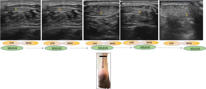
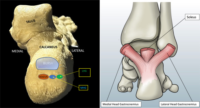
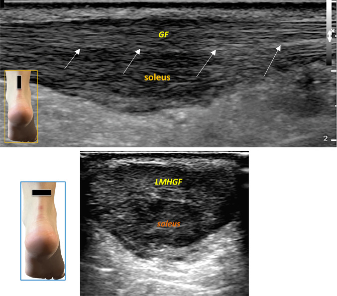
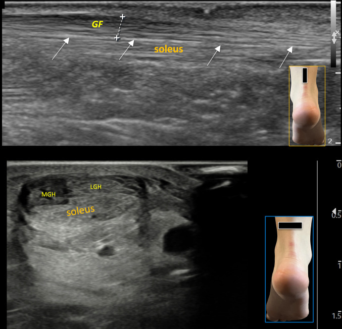
This pictorial essay clarifies the complex anatomical and ultrasonographic features of the Achilles tendon. Utilizing high-frequency ultrasound probes (12-18 MHz), the study visualizes the tripartite segmentation of the tendon as it relates to anatomical fascicles. Notably, there is variability in the ultrasound visibility of fascicles and this partition influences biomechanical properties and susceptibility to injury. This essay not only enhances the understanding of Achilles tendon anatomy but also underscores the importance of anatomical knowledge for comprehending associated pathologies. The findings suggest potential areas for further research into tendon pathology, facilitating the development of targeted rehabilitation strategies.

Keywords: Achilles tendon; Achilles tendon fasciculation; Retrocalcaneal bursa; Tendinopathy; Ultrasound.


Conflict of interest statement

Declarations. Conflict of interest: The authors declare that they have no known competing financial interests or personal relationships that could have appeared to influence the work reported in this paper. Ethical approval: Ethical approval is not required for this work. Consent: Not applicable. Registration of research studies: Not applicable.

Figures

Fig. 1
Fig. 1
A Longitudinal scan of the Achilles tendon showing its two components: the superficial gastrocnemius fascicle, the deep soleus fascicle and the septum in the middle. Below the figure, a schematic illustration of the anatomical section is shown. B Axial scan of the Achilles tendon. Note the three components, two in the upper portion (gastrocnemius) and one in the lower part (soleus). The boundary between the two heads of the gastrocnemius appears indistinct on short-axis ultrasound, and no clear vertical septum is visible. The drawing under this figure illustrates the anatomical section. C Schematic anatomical transverse section of the Achilles tendon and its regional anatomical relationships. LMHGF Lateral and Medial Head of Gastrocnemius Fascicles; FHL Flexor Hallucis Longus muscle, PT Peroneal Tendons, FDL Flexor Digitorum Longus tendon, PTT Posterior Tibial Tendon
Fig. 2
Fig. 2
A In this case of moderate tendinopathy, the longitudinal scan of the Achilles tendon shows peritenon duplication of the septum, with the two fascicles maintaining their own peritenon. B Enlarged detail (in long-axis scan) of the dashed rectangular area shown in A. White arrows indicate the double peritenon. C The separation of the fascicles with peritenon duplication is also clearly visible in axial section. A small amount of effusion is visible between the two layers of the duplicated peritenon. White arrows indicate the double peritenon
Fig. 3
Fig. 3
In long-axis scans, the two fascicles may occasionally have a distinct anatomical boundary due to separate insertions on the heel bone. In such cases, the soleus tendon is clearly separated from the gastrocnemius fascicle, inserting into the calcaneus with a fat triangle between the two components. This anatomical variant is present in monkeys. The figure shows divergent yellow dotted lines representing the soleus and gastrocnemius fascicles (GHF) inserting separately into the heel
Fig. 4
Fig. 4
Long-axis scan of the Achilles tendon showing the accessory soleus muscle, which has its own muscle belly and tendon that inserts anteriorly and separately to the Achilles tendon enthesis. FHL Flexor Hallucis Longus. The green arrows indicate the belly and tendon of the accessory soleus, which inserts into the heel
Fig. 5
Fig. 5
Schematic anatomical drawing showing the triceps surae fascicles crossing each other. The white dash-dotted line represents the direction of the gastrocnemius fascicles, while the black dashed line represents the direction of the soleus fascicle. The box on the right is an enlargement of the drawing. MHG Medial Head of Gastrocnemius, LHG Lateral Head of Gastrocnemius, LMHGF Lateral and Medial Head of Gastrocnemius, LAT MALL lateral malleolus, MED MALL medial malleolus
Fig. 6
Fig. 6
Short-axis scan of the three components of the left Achilles tendon in a dynamic sequence from top (starting with the first figure on the left) to bottom (last figure on the right). S soleus fascicle, LHG Lateral Head of Gastrocnemius, MHG Medial Head of Gastrocnemius
Fig. 7
Fig. 7
on the left, the insertions of the three fascicles into the calcaneal tuberosity on the calcaneus bone (see text). LHG Lateral Head of Gastrocnemius, MHG Medial Head of Gastrocnemius, BURSA retrocalcaneal bursa. On the right, schematic drawing of the three fascicles inserting into the heel
Fig. 8
Fig. 8
Evidence of fasciculations becomes more apparent in tendinopathy, where often only one partition (more frequently the soleus fascicle) is affected, while the remaining portion remains uninvolved, exhibiting a normal echopattern or mild pathological involvement. The figure shows a long-axis scan (above) of the Achilles tendon, predominantly affecting the soleus fascicle. GF Gastrocnemius fascicle. White arrows show the septum between the two components. The lower figure shows a short-axis scan: LMHGF Lateral and Medial Head of Gastrocnemius Fascicles
Fig. 9
Fig. 9
In tendinopathy, it is easier to distinguish the fascicles, identifying which partition is most affected. In longitudinal scans, the tendon appears bifasciculate, while in axial scans, a tripartite pattern is observed. In the upper figure, tendinopathy starts to affect the superficial layer (GF gastrocnemius fascicle). White arrows indicate the septum between the two components. The lower figure shows another case of focal tendinopathy affecting only the medial head of the gastrocnemius (MHG) in a short-axis scan, with the LHG (lateral head of gastrocnemius) appearing normal
Fig. 10
Fig. 10
In cases of chronic tendinopathy, longitudinal fissures (intramural lesions) can also be identified, where partial tears localize in the septum between the gastrocnemius and soleus bundles. The Achilles tendon is shown between the calipers, with white arrows marking the fissures
Fig. 11
Fig. 11
Dashed yellow arrows show the movement of the tongue-like process of the distal Kager’s fat pad (T1, T2) during the Thompson test. In this subject, the tendon elevates as the adipose process enters the bursal space. It has been hypothesized that this process may have a trophic and protective function for the overlying tendon. The schematic anatomical drawing below shows the position of Kager’s fat pad in relation to the Achilles tendon and the tongue-like process inside the bursa. AT Achilles tendon; the white arrowhead indicates the elevation of the tendon due to the movement of Kager's tongue-like process
Fig. 12
Fig. 12
A short-axis scan of the fascicles slightly proximal to the enthesis, the soleus is directed on the medial surface of the calcaneal tuberosity (slightly superiorly) and both the two component of gastrocnemius are in lateral position. In this preinsertional scan the two fascicles of the gastrocnemius appear as a single fascicle. The figure below (great white arrow) is the same of the above one, with the indication for the two fascicles (soleus with red dots and both the fascicles of gastrocnemius with green dots). MED medial, LAT lateral. GF gastrocnemius fascicle. B short-axis scan at the insertion of the two fascicles of gastrocnemius, slight lateral and distally at the previous scan: the figure below (great white arrow) is the same of the above one, with the indication for the two fascicles of gastrocnemius: the lateral head of gastrocnemius (LHG, green dots) is inserted on the lateral surface of the calcaneal tuberosity and the medial head (MHG, blue dots), adjacent and medially to it. The soleus fascicle (not in figure) is inserted on the medial surface of the calcaneal tuberosity but slightly more superiorly. Between the two figures, there’s a schematic drawing (framed by a white line) of the three fascicles inserting into the heel

References

    1. Wood Jones F. Structure and function as seen in the foot. London: Bailliere, Tindall and Cox, 1944:124–133.
    1. Szaro P, Witkowski G, Smigielski R, Krajewski P, Ciszek B (2009) Fascicles of the adult human Achilles tendon—an anatomical study. Ann Anat 191(6):586–593 - PubMed
    1. Ballal MS, Walker CR, Molloy AP. The anatomical footprint of the Achilles tendon: a cadaveric study. Bone Joint J. 2014 Oct;96-B(10):1344–8. - PubMed
    1. Stella SM, Ciampi B, Caviglia e Piede richiami di anatomia e semeiotica ecografica; Atlante di Anatomia Ecografica e Biomeccanica Muscoloscheletrica. Ed. Piccin Padova, 2017. Pagg 543–545.
    1. Chiarugi G, Bucciante L, Istituzioni di Anatomia dell’Uomo, Vol2, App. Muscolare, Muscolo Soleo, Cap 4, Pag 247. Ed. Vallardi, Milano, XI edizione 1977.

MeSH terms

LinkOut - more resources