. 2025 Feb 3;21(2):e1012857.
doi: 10.1371/journal.ppat.1012857. eCollection 2025 Feb.

Iron-mediated post-transcriptional regulation in Toxoplasma gondii

Affiliations

Iron-mediated post-transcriptional regulation in Toxoplasma gondii

Megan A Sloan et al. PLoS Pathog. .

Abstract

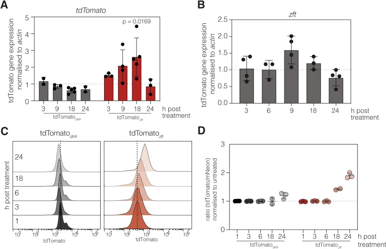
Iron is required to support almost all life; however, levels must be carefully regulated to maintain homeostasis. Although the obligate parasite Toxoplasma gondii requires iron, how it responds upon iron limitation has not been investigated. Here, we show that iron depletion triggers significant transcriptional changes in the parasite, including in iron-dependent pathways. We find that a subset of T. gondii transcripts contain stem-loop structures, which have been associated with post-transcriptional iron-mediated regulation in other cellular systems. We validate one of these (found in the 3' UTR of TGME49_261720) using a reporter cell line. We show that the presence of the stem-loop-containing UTR is sufficient to confer accumulation at the transcript and protein levels under low iron. This response is dose and time-dependent and is specific for iron. The accumulation of transcript is likely driven by an increased reporter mRNA stability under low iron. Interestingly, we find iron-mediated changes in mRNA stability in around 400 genes. To examine the potential mechanism of this stability, we tested aconitase interaction with mRNA in low iron and found 43 enriched transcripts, but no specific interaction with our reporter UTR. However, the endogenous UTR led to maintenance of protein levels and increased survival of the parasite under low iron. Our data demonstrate the existence of iron-mediated post-transcriptional regulation in Toxoplasma for the first time; and suggests iron-mediated regulation may be important to the parasite in low iron environments.


Conflict of interest statement

The authors have declared that no competing interests exist.

Figures

Fig 1
Fig 1. The T. gondii transcriptional response to iron deprivation.
A. Volcano plot from RNAseq data comparing untreated ΔKu80 to parasites cultured in 100 µM DFO for 24 hours. Adjusted p-values from the Wald test with Benjamini and Hochberg correction. Cut-offs shown with dashed lines are p-adj < 0.05 and log2 fold change of >2 or <−2. B. Venn diagram showing overlap between significantly regulated genes from DFO-treated parental and ΔBFD1 parasites. C. Heatmap from RNAseq dataset depicting z-scores of genes shown to be regulated by BFD1 (Waldman et al. 2020) [35], in parental and ΔBFD1, DFO treated and untreated parasites. D. Schematic showing the filtering process to identity transcripts containing IRE-like sequences in the T. gondii transcriptome. E. Volcano plot as in (A) where transcripts with IRE-like sequences in classes I (red), II (orange) and VIII (gold) are highlighted. See text for more details.
Fig 2
Fig 2. The zft 3’ UTR is sufficient to render a reporter gene iron responsive.
A. Schematic of the tdTomatodhfr cassette and tdTomatozft cassette. Both reporters were integrated into the uprt locus in a ΔKu80:mNeonGreen parasite line. B. Contour plots of mNeonGreen and tdTomato expression in untreated and treated with 100 µM DFO for 24 hours (measured by flow cytometry) for both reporter lines. Representative of at least 5000 parasites from a single experiment. C. Plot showing the mean tdTomato:mNeonGreen fluorescence ratio in tdTomatodhfr and tdTomatozft parasites treated with either 100 µM DFO or 2.5 mM ferric ammonium citrate (FAC), normalised to untreated parasites. Plot shows data from 5 independent experiments. p values compared to tdTomatodhfr by one way ANOVA with Dunnett’s correction D. Immunofluorescence showing mNeonGreen and tdTomato expression in untreated and DFO treated (24 hours, 100 µM DFO) tdTomatodhfr parasites with a violin/box plot showing the quantification of tdTomato:mNeonGreen fluorescence from vacuoles from two biological replicates (n indicated on graph), p values from unpaired t test. Scale bar 5 µm E. Immunofluorescence showing mNeonGreen and tdTomato expression in untreated and DFO treated (24 hours, 100 µM DFO). Scale bar 5 µm. tdTomatozft parasites with a violin/box plot showing the quantification of tdTomato:mNeonGreen ratio as above. F. Representative overlapping histograms showing tdTomato fluorescence in the tdTomatoisu1 reporter line both untreated and after 24 hours of treatment in 100 µM DFO. G. Plot showing the mean tdTomato:mNeonGreen fluorescence ratio in untreated tdTomatoisu1 and those treated with 100 µM DFO, normalised to untreated parasites. Data shown from 4 independent experiments. p value from unpaired t test. H. Histogram showing the frequency of differentially regulated transcripts with IRE-like sequences in their 3’ UTRs in RNAseq data. Transcripts were binned (bin size = 400 bp) based on distance of the IRE-like sequence from the stop codon.
Fig 3
Fig 3. ZFT responsiveness is dose dependent and specific to iron chelation.
A. Representative overlapping histograms showing tdTomato fluorescence in the tdTomatodhfr and tdTomatozft reporter lines treated with indicated DFO concentration for 24 hours. B. Quantification of change of tdTomato:mNeonGreen ratio. p values from one way ANOVA with Dunnett’s correction, compared to tdTomatodhfr + 500 µM DFO C. Quantification in luminescence signal (normalised to parasite flourescense and untreated cells) after treatment with indicated DFO concentration for 24 h. p values from one way ANOVA with Dunnett’s correction, compared to parasites treated with 0.5 µM DFO D. Representative overlapping histograms showing tdTomato fluorescence, measured by flow cytometry, in the tdTomatodhfr and tdTomatozft reporter lines treated with 100 µM DFO, 100 µM BIP or 100 µM PIH for 24 hours. E. Mean tdTomato:mNeonGreen fluorescence ratio for three independent experiments. p values from one way ANOVA with Dunnett’s correction, compared to tdTomatodhfr + DFO. F. Fluorescence growth assay for tdTomato parasites cultured in increasing concentrations of iron chelators. Points show mean of three independent experiments ± SEM. G. Representative overlapping histograms showing tdTomato fluorescence, measured by flow cytometry, in the tdTomatodhfr and tdTomatozft reporter lines treated with 100 µM DFO, 5 µM TPEN (Zn chelator) or 25 µM TTM (Cu chelator) for 24 hours. H. Mean tdTomato:mNeonGreen fluorescence ratio for three independent experiments. p values from one way ANOVA with Dunnett’s correction, compared to tdTomatodhfr + DFO.
Fig 4
Fig 4. Kinetics of tdTomatozft iron-responsiveness.
qRT-PCR on the relative abundance of tdTomato (A) and zft (B) transcripts (normalised to actin) in the tdTomatodhfr and tdTomatozft reporter lines after treatment with 100 µM DFO, compared to untreated parasites for indicated time. Points represent a single experiment, bars at mean ± SD. C. Representative overlapping histograms showing tdTomato fluorescence, measured by flow cytometry, in the tdTomatodhfr and tdTomatozft reporter lines treated with 100 µM DFO over a 24-hour time course. D. Plot showing the mean tdTomato:mNeonGreen fluorescence ratio for the experiments described in (C).
Fig 5
Fig 5. Removal of the IRE-like element from the zft 3’ UTR reduces iron responsiveness.
A. Schematic of the iron reporter cassettes integrated into the uprt locus. B. qRT-PCR on the relative abundance of tdTomato transcripts (normalised to actin) in the tdTomatodhfr, tdTomatozft and tdTomatozftΔIRE reporter lines after 18 hours treatment with 100 µM DFO, compared to untreated parasites. Bars at mean ± SD, p values from one way ANOVA with Tukey’s correction. Bars at mean ± SD, p values from one way ANOVA with Tukey’s correction. C. Representative overlapping histograms showing tdTomato fluorescence in the tdTomatodhfr, tdTomatozft and tdTomatozftΔIRE reporter lines, treated with 100 µM DFO for 24 hours. D. Mean tdTomato:mNeonGreen fluorescence ratio, p values from one way ANOVA with Dunnett’s correction, compared to tdTomatodhfr + DFO. E. Representative overlapping histograms showing tdTomato fluorescence in the tdTomatodhfr, tdTomatozft and tdTomatozftΔIRE reporter lines, treated with alkaline stress for 24 hours. D. Mean tdTomato:mNeonGreen fluorescence ratio, p values from one way ANOVA with Dunnett’s correction, compared to tdTomatodhfr + stress.
Fig 6
Fig 6. zft 3’UTR demonstrates iron-dependent mRNA stability.
Untreated or DFO-treated tdTomatodhfr (A) or tdTomatozft (B) or parental parasites (C) treated intracellularly with actinomycin D and qPCR performed for indicated gene. Results normalised to actin, results mean of three independent replicates, ± SD, p value from extra sum of squares F test. D. mRNA stability performed as above, targeting sag1, Tg500296, Tg253510 or isu1, normalised to actin. Results mean of three independent replicates, ± SD, p values from extra sum of squares F test, due to the lack of degradation for isu1, we were unable to calculate a p value. E. Average abundance for transcripts which reached <75% of t = 0 across a 5-hour time course of actinomycin D treatment from untreated parasites. Transcripts were binned based on decay kinetics. Bin 1- 50% abundance by 3 hours, then further decay; Bin 2 - 50% abundance by 3 hours, without further decay and Bin 3 – transcripts which did not decay >50% during the time course F. Binned average transcripts remaining from DFO-treated parasites, normalised to parasites cultured in standard conditions. Binning information above. G. Selected motifs significantly enriched in DFO-responsive transcripts (from XSTREME), with proportion of transcripts containing the motif indicated. H. Schematic showing protocol for aconitase IPs. I. Blots showing the immunoprecipitation of mNeonGreen-Ty and ACN-Ty from T. gondii parasites. CDPK1 included as cytosolic control. Wash – the output of the 6th and final washing step. Representative of three independent replicates. DNA-agarose gel showing qPCR products amplified from reverse transcribed RNA from either starting lysates or RNA co-precipitated with mNeonGreen-Ty and aconitase-Ty proteins. PCR amplicons for actin (expected size: 188 bp), zft (expected size: 116 bp) and rng2 (expected size: 146 bp). J. qRT-PCR results from IPs performed in (H). Points represent average Ct values from individual biological replicates, performed in triplicate, lines at median. ND – not detected.
Fig 7
Fig 7. Presence of zft 3’ UTR affects native protein.
A. Blots showing ZFT-HAsag1 protein levels after 18 and 24 h treatment with DFO. Protein levels are significantly (t test, compared to untreated) reduced at both time points. Bars at mean ± SD, points represent values from independent biological experiments. B. ZFT-HAzft parasites tagged with the native UTR showed significant reduction at 24 h of treatment, but protein levels were maintained at 18 h. C. Host cell monolayer absorbance after infection in the presence of increasing DFO. Points show mean of four independent experiments ± SEM. D. Representative plaque assays after 7 days of growth either untreated or in the presence of 1 µM DFO. E. Plaque area quantification showing % change in plaque area compared to untreated. Bars at mean ± SD and points represent mean plaque area from four independent experiments. p values from one way ANOVA with Tukeys correction.

Similar articles

References

    1. Bergmann A, Floyd K, Key M, Dameron C, Rees KC, Thornton LB, et al.. Toxoplasma gondii requires its plant-like heme biosynthesis pathway for infection. PLOS Pathog. 2020;16:e1008499. doi: 10.1371/journal.ppat.1008499 - DOI - PMC - PubMed
    1. Pamukcu S, Cerutti A, Bordat Y, Hem S, Rofidal V, Besteiro S. Differential contribution of two organelles of endosymbiotic origin to iron-sulfur cluster synthesis and overall fitness in Toxoplasma. PLoS Pathog. 2021;17(11):e1010096. doi: 10.1371/journal.ppat.1010096 - DOI - PMC - PubMed
    1. Sloan MA, Aghabi D, Harding CR. Orchestrating a heist: uptake and storage of metals by apicomplexan parasites. Microbiology (Reading). 2021;167(12):mic.0.001114. doi: 10.1099/mic.0.001114 - DOI - PMC - PubMed
    1. Aghabi D, Sloan M, Gill G, Hartmann E, Antipova O, Dou Z, et al.. The vacuolar iron transporter mediates iron detoxification in Toxoplasma gondii. Nat Commun. 2023;14(1):3659. doi: 10.1038/s41467-023-39436-y - DOI - PMC - PubMed
    1. Reinert A, Morawski M, Seeger J, Arendt T, Reinert T. Iron concentrations in neurons and glial cells with estimates on ferritin concentrations. BMC Neurosci. 2019;20(1):25. doi: 10.1186/s12868-019-0507-7 - DOI - PMC - PubMed