. 2025 Feb 3;16(1):1283.
doi: 10.1038/s41467-025-56491-9.

Structural insights into light-gating of potassium-selective channelrhodopsin

Affiliations

Structural insights into light-gating of potassium-selective channelrhodopsin

Takefumi Morizumi et al. Nat Commun. .

Abstract

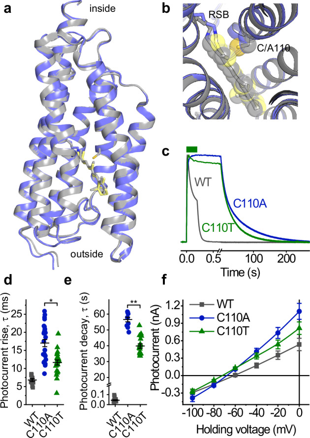
Structural information on channelrhodopsins' mechanism of light-gated ion conductance is scarce, limiting its engineering as optogenetic tools. Here, we use single-particle cryo-electron microscopy of peptidisc-incorporated protein samples to determine the structures of the slow-cycling mutant C110A of kalium channelrhodopsin 1 from Hyphochytrium catenoides (HcKCR1) in the dark and upon laser flash excitation. Upon photoisomerization of the retinal chromophore, the retinylidene Schiff base NH-bond reorients from the extracellular to the cytoplasmic side. This switch triggers a series of side chain reorientations and merges intramolecular cavities into a transmembrane K+ conduction pathway. Molecular dynamics simulations confirm K+ flux through the illuminated state but not through the resting state. The overall displacement between the closed and the open structure is small, involving mainly side chain rearrangements. Asp105 and Asp116 play a key role in K+ conductance. Structure-guided mutagenesis and patch-clamp analysis reveal the roles of the pathway-forming residues in channel gating and selectivity.


Conflict of interest statement

Competing interests: The authors declare no competing interests.

Figures

Fig. 1
Fig. 1. Cryo-EM density map and structure of HcKCR1_C110A trimer embedded in peptidisc in the dark-adapted state (left) and laser-flash-illuminated state (right).
a Peptidisc comprising a C3-symmetric HcKCR1_C110A trimer in the dark (blue, magenta, and orange) and upon laser-flash illumination (green, pink, and yellow), lipids and cholesterols (gray), and multiple surrounding 37-residue amphipathic peptides (white). View from the intracellular side. b Structure models of dark-adapted state (left) and illuminated state (right) HcKCR1_C110A viewed from the intracellular side (upper) and the membrane plane (lower) with all-trans-retinal (yellow) and 13-cis retinal (red), respectively. In one protomer of the trimer, the helices are labeled to show the order of transmembrane helices.
Fig. 2
Fig. 2. Comparison of the HcKCR1_C110A and wild-type HcKCR1 (PDB: 8GI8) dark-adapted structures and functional characterization of C110 mutants.
a An overlay of structures of dark-adapted C110A mutant (shown in blue, retinal in yellow) and wild-type (WT, shown in gray). The helices are shown as cartoons, the chromophore and the residue 110 side chains, as sticks. b A zoom-in into the residue 110 region. c Representative photocurrent traces recorded at 0 mV in response to 200-ms light pulses (green bar) normalized at the peak value. d, e Photocurrent rise and decay time constants. The symbols are individual-cell data; the lines are mean ± sem values. *p = 3.7E-4 (n = 10, 26, and 23 cells for the WT, C110A, and C110T, respectively); **p = 4.3E-5 (n = 16, 10, and 17 cells for the WT, C110A, and C110T, respectively) by the two-tailed Mann–Whitney test. f Current-voltage dependencies (mean ± sem, n = 25, 37, and 40 cells for the WT, C110A, and C110T, respectively). Source data are provided as a Source Data file.
Fig. 3
Fig. 3. Intramolecular cavities merge upon illumination to form an aqueous cation conduction pathway.
a Cavities in HcKCR1_C110A protomer modeled with the HOLLOW software. The cavity color shows the electrostatic potential from −5 (red) to +5 (blue). The helices are shown as cartoons; the chromophore, as a stick model. b The CAVER software detects an intramolecular tunnel (yellow) only in the laser-flash-illuminated HcKCR1_C110A structure, not in the dark-adapted state structure. c MD simulations show a water chain connecting the RSB with the intracellular aqueous phase in the structure of the laser-flash-illuminated HcKCR1_C110A trimer.
Fig. 4
Fig. 4. The three segments of the cation conduction path.
Structures of dark-adapted and laser-flash-illuminated HcKCR1_C110A containing cavities modeled with HOLLOW; the helices are shown as cartoons; the chromophore and key residues, as stick models; water molecules, as small spheres; and the potassium ions transported in molecular dynamics simulations (right panel), as large green spheres.
Fig. 5
Fig. 5. Mutational analysis on the tunnel constricting residues identified in the structure of laser-flash-illuminated HcKCR1_C110A.
a A tunnel profile detected by CAVER using the probe radius 1 Å. C1-C4 are the four constrictions. The lines show the positions of the tested residues along the tunnel. b, Photocurrent traces recorded from the wild-type (WT) and Y222T mutant at incremental voltages. c The current-voltage relationships of the WT and Y222T mutant. The symbols are mean ± sem (n = 19 and 17 cells, respectively). d The Vr values are derived by analysis of the current-voltage dependencies (see Methods for solution compositions). e Photocurrent amplitudes normalized to the WT recorded in the same experiment. d, e The symbols are individual-cell data recorded at the time of peak (empty circles) and the end of illumination (filled circles); the lines are mean ± sem values. *p < 0.05; **p < 0.001; ***p < E-5 compared to the WT by the one-way ANOVA with the Tukey means comparison. The numbers of the cells sampled for each variant and the exact p values are provided in the Source Data file.

References

    1. Deisseroth, K. Optogenetics: 10 years of microbial opsins in neuroscience. Nat. Neurosci.18, 1213–1225 (2015). - PMC - PubMed
    1. Emiliani, V. et al. Optogenetics for light control of biological systems. Nat. Rev. Method Primers2, 55 (2022). - PMC - PubMed
    1. Piatkevich, K. D. & Boyden, E. S. Optogenetic control of neural activity: the biophysics of microbial rhodopsins in neuroscience. Q. Rev. Biophys.57, e1 (2023). - PubMed
    1. Feldbauer, K. et al. Channelrhodopsin-2 is a leaky proton pump. Proc. Natl. Acad. Sci. USA106, 12317–12322 (2009). - PMC - PubMed
    1. Govorunova, E. G., Sineshchekov, O. A., Janz, R., Liu, X. & Spudich, J. L. Natural light-gated anion channels: A family of microbial rhodopsins for advanced optogenetics. Science349, 647–650 (2015). - PMC - PubMed

MeSH terms