. 2025 Feb 3;16(1):1276.
doi: 10.1038/s41467-025-56612-4.

Structural insights into dynamics of the BMV TLS aminoacylation

Affiliations

Structural insights into dynamics of the BMV TLS aminoacylation

Wen Yang et al. Nat Commun. .

Abstract

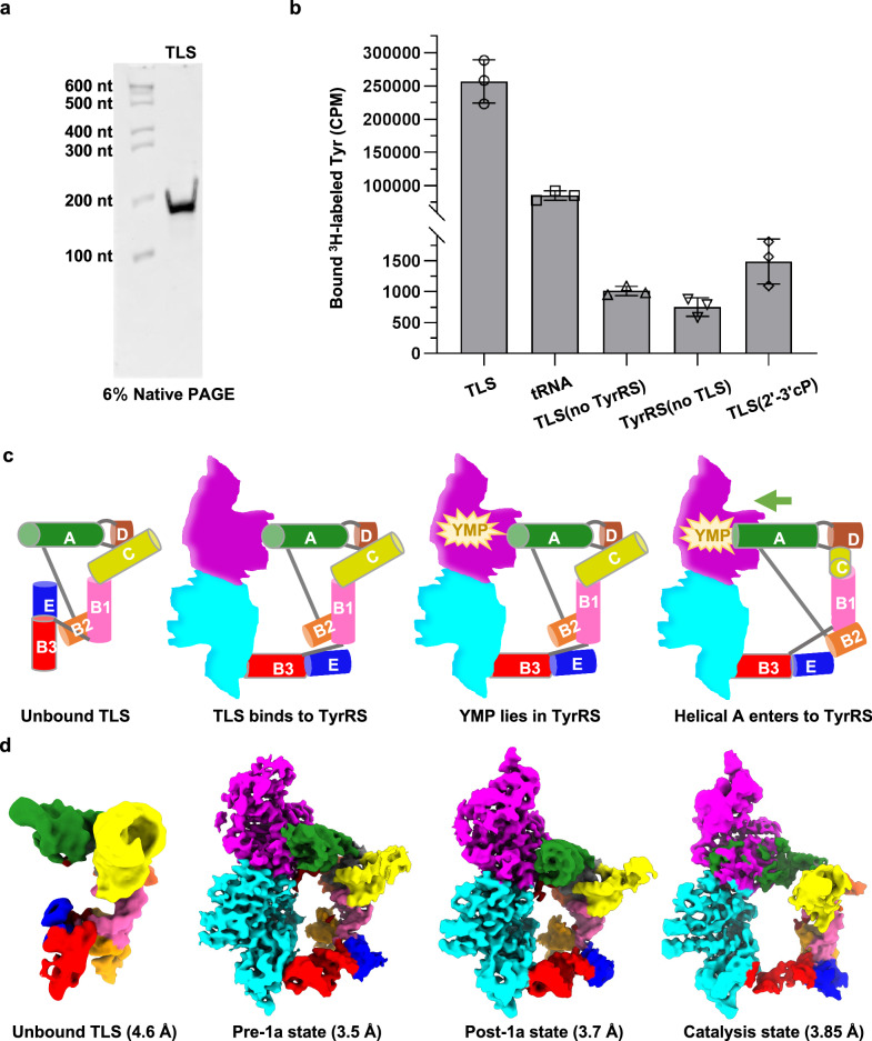
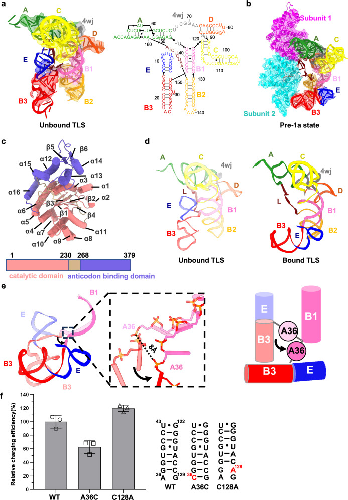
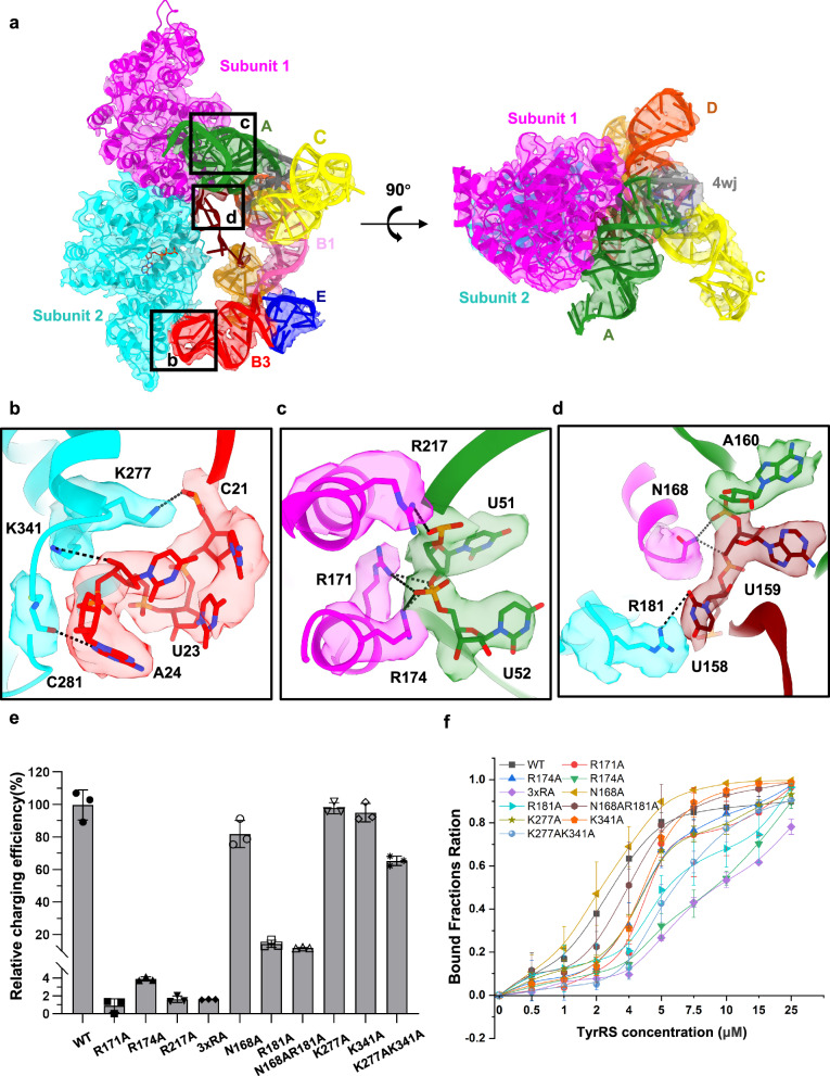
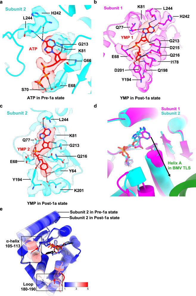
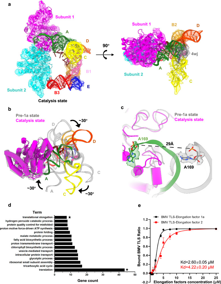
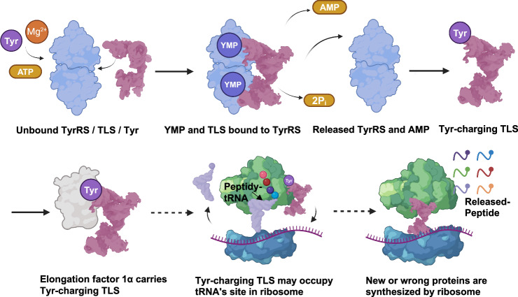
Brome Mosaic Virus (BMV) utilizes a tRNA-like structure (TLS) within its 3' untranslated region to mimic host tRNA functions, aiding aminoacylation and viral replication. This study explores the structural dynamics of BMV TLS interacting with tyrosyl-tRNA synthetase (TyrRS) during aminoacylation. Using cryo-EM, we capture multiple states of the TLS-TyrRS complex, including unbound TLS, pre-1a, post-1a, and catalysis states, with resolutions of 4.6 Å, 3.5 Å, 3.7 Å, and 3.85 Å, respectively. These structural comparisons indicate dynamic changes in both TLS and TyrRS. Upon binding, TLS undergoes dynamic rearrangements, particularly with helices B3 and E pivoting, mediated by the unpaired A36 residue, ensuring effective recognition by TyrRS. The dynamic changes also include a more compact arrangement in the catalytic center of TyrRS and the insertion of 3' CCA end into the enzyme's active site, facilitating two-steps aminoacylation. Enzymatic assays further demonstrated the functional importance of TLS-TyrRS interactions, with mutations in key residues significantly impacting aminoacylation efficiency. Furthermore, Electrophoretic Mobility Shift Assay (EMSA) demonstrated that BMV TLS binds elongation factors EF1α and EF2, suggesting a multifaceted strategy to exploit host translational machinery. These findings not only enhance our knowledge of virus-host interactions but also offer potential targets for antiviral drug development.


Conflict of interest statement

Competing interests: The authors declare no competing interests.

Figures

Fig. 1
Fig. 1. Cryo-EM analysis of BMV TLS-TyrRS during the aminoacylation reaction.
a Native PAGE demonstrating the high conformational homogeneity of BMV TLS. More than 3 times this experiment was repeated independently with similar results. b In vitro aminoacylation assay comparing the activity of TyrRS with TLS and tRNA (both at the same final concentration). Data are shown as the mean ± SD (n = 3 independent experiments). Source data are provided as a Source Data file. This assay includes three controls: “TLS (no TyrRS)” indicates a reaction system without the addition of TyrRS; “TyrRS (no TLS)” indicates a reaction system without BMV TLS; and “TLS (2′–3′ cP)” refers to TLS where the 3′ terminal adenine is circularized into a 2′–3′ adenosine cyclic phosphate, which prevents aminoacylation at the 2′-OH group of the ribose. c Schematic illustration of the aminoacylation process catalyzed by TyrRS. The differently colored cylinders represent various TLS helices; gray lines between cylinders indicate the connections between helices; Magenta and cyan blocks represent the two subunits of TyrRS; the yellow star represents YMP; and the green arrow indicates the movement of TLS Helix A. d Cryo-EM maps showing various conformations of TLS-TyrRS engaged in aminoacylation. The maps correspond to the states illustrated in (c), from left to right.
Fig. 2
Fig. 2. The unpaired residue A36-mediated dynamic pivoting of Helices E and B3 is crucial for the efficient recognition by TyrRS.
a Schematic representation of the unbound TLS structure. The left panel shows the cryo-EM map and atomic model of the unbound TLS, while the right panel illustrates its secondary structure. b Superimposed model and map of TLS-TyrRS in the pre-1a state. c Domain organization of a single TyrRS subunit. d Comparison of the unbound TLS structure (left, transparent with colors) and the bound TLS structure (right, solid colors), demonstrating conformational changes upon binding. e Overlay of the B1, B3, and E helices in the two states of TLS, along with a magnified view of the detailed alignment at residue A36 (transparent colors for unbound TLS, solid colors for bound TLS). Helix B1 remains almost unchanged, while A36, located at the junction of helices B1 and B3, shows an 8-Å displacement in the phosphate backbone. A cartoon schematic (right) illustrates how A36 mediates the dynamic rearrangement of helices E and B3. f In vitro aminoacylation assay to confirm the importance of A36. Data are shown as the mean ± SD (n = 3 independent experiments). Source data are provided as a Source Data file.
Fig. 3
Fig. 3. Binding interfaces between BMV TLS and TyrRS in the pre-1a state.
a Superimposed model and map of the pre-1a state, showing three sets of interaction interfaces. Black boxes indicate the regions will be shown in (bd). b Detailed interactions between the TLS’s B3 domain and the anticodon-binding domain of TyrRS subunit 2. c Detailed interactions between the TLS’s A domain and the catalytic domain of TyrRS subunit 1. Black dashed lines indicate hydrogen bonds. d Detailed interactions between the TLS’s wobble loop and the TyrRS’s catalytic domains. e Validation of key residues for TyrRS’s enzymatic activity with TLS. “3xRA” represents the combined mutations of R171A, R174A, and R217A. Data are shown as the mean ± SD (n = 3 independent experiments). Source data are provided as a Source Data file. (f) Validation of key residues for TyrRS’s binding affinity with TLS. Data are shown as the mean ± SD (n = 3 independent experiments). Source data are provided as a Source Data file.
Fig. 4
Fig. 4. ATP/YMP Pocket in TyrRS.
a ATP-binding pocket in the subunit 2 of TyrRS in the pre-1a state, showing detailed interactions between ATP and key residues. Black dashed lines represent hydrogen bonds. b YMP-binding pocket in the subunit 1 of TyrRS in the post-1a state (YMP1). Detailed interactions between YMP1 and key residues are shown, with black dashed lines representing hydrogen bonds. c YMP-binding pocket in the subunit 2 of TyrRS in the post-1a state (YMP2). d Comparison between the YMP-binding pockets in the post-1a state. The black arrow indicates that YMP1 is positioned closer to the TLS acceptor arm (forest green) compared to YMP2. e Comparison of the subunit 2’s catalytic domain between the pre-1a state (transparent colors) and post-1a state (solid colors), colored by C-alpha RMSD in ChimeraX. ATP from the pre-1a state is shown in gray, and YMP2 from the post-1a state is shown in black. The black dashed boxes highlight the regions with the most significant structural changes, specifically α-helix 105–113 and loop 180–190. Red arrows indicate the direction of movement, with the catalytic domain of subunit 2 in the post-1a state shifting closer to the active site.
Fig. 5
Fig. 5. Structural arrangements in the BMV TLS during the second-step aminoacylation by TyrRS.
a Superimposed models and maps of the catalysis state. b Comparison between the pre-1a state (gray) and the catalysis state (colored). TLS rotates approximately 30° around TyrRS, with the black arrow indicating the direction of rotation. c Close-up structural comparison between the pre-1a state (gray) and the catalysis state (colored). The black dashed line represents the distance between the phosphate backbones of residue A169 at the 3′ end of the TLS acceptor arm in both states, measuring approximately 29 Å. d Top 15 of GO Enrichment. Black asterisks highlight translation and translational elongation Biological Processes (GOTERM-BP). Biotin-antisense TLS is control group. Data are shown as the mean (n = 2 independent experiments). Source data are provided as a Source Data file. e Binding affinity of BMV TLS-EF1α and BMV TLS-EF2. Binding curves were generated from EMSA experiments (n = 3 independent experiments). Data are shown as the mean ± SD. Source data are provided as a Source Data file.
Fig. 6
Fig. 6. Cartoon illustration of TyrRS-mediated TLS aminoacylation reaction and TLS interactions with elongation factors to harness host translational machinery.
Upon binding with TyrRS, BMV TLS undergoes significant conformational rearrangements, which allow it to be recognized by TyrRS to charge with Tyr, mimicking a tRNA function. The Tyr-charging TLS then binds with EF1α or EF2 to manipulate the host’s translational machinery to its advantage. Created in BioRender. N′N′, OY. (2025) https://BioRender.com/q47d213.

Similar articles

References

    1. Kao, C. C. & Sivakumaran, K. Brome mosaic virus, good for an RNA virologist’s basic needs. Mol. Plant Pathol.1, 91–97 (2000). - PubMed
    1. Rao, A. L. N. & Cheng Kao, C. The brome mosaic virus 3’ untranslated sequence regulates RNA replication, recombination, and virion assembly. Virus Res.206, 46–52 (2015). - PubMed
    1. Rietveld, K., Pleij, C. W. & Bosch, L. Three-dimensional models of the tRNA-like 3’ termini of some plant viral RNAs. EMBO J.2, 1079–1085 (1983). - PMC - PubMed
    1. Sherlock, M. E., Hartwick, E. W., MacFadden, A. & Kieft, J. S. Structural diversity and phylogenetic distribution of valyl tRNA-like structures in viruses. RNA27, 27–39 (2021). - PMC - PubMed
    1. Matsuda, D. & Dreher, T. W. The tRNA-like structure of Turnip yellow mosaic virus RNA is a 3’-translational enhancer. Virology321, 36–46 (2004). - PubMed

LinkOut - more resources