. 2025 Feb 3;15(1):4035.
doi: 10.1038/s41598-025-88621-0.

Establishment of a bat lung organoid culture model for studying bat-derived infectious diseases

Affiliations

Establishment of a bat lung organoid culture model for studying bat-derived infectious diseases

Mohamed Elbadawy et al. Sci Rep. .

Abstract

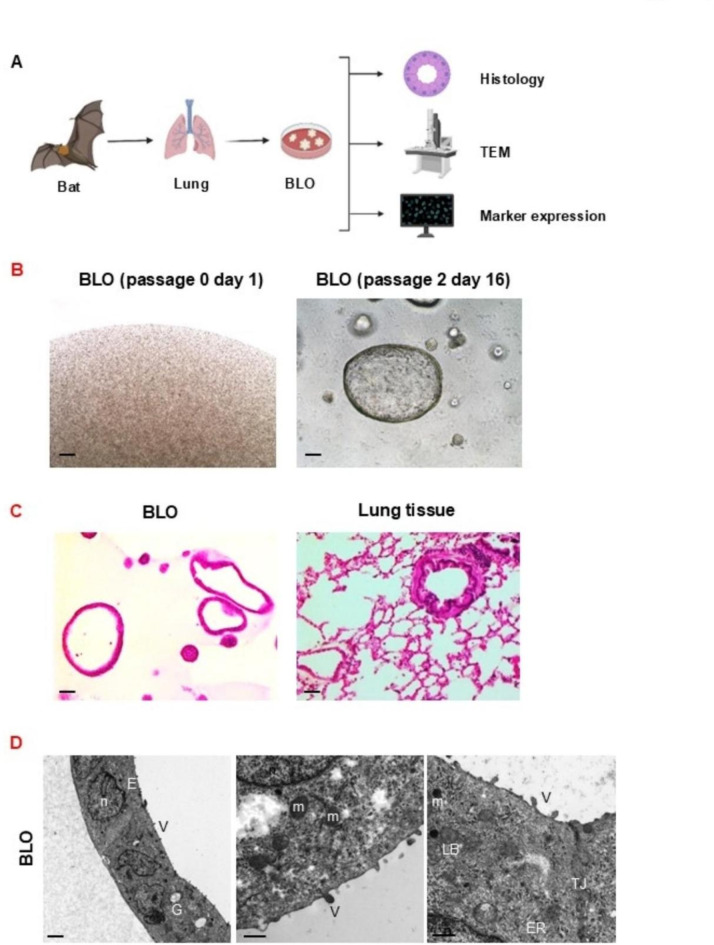
Bat is considered a natural reservoir of various important pathogens, including severe acute respiratory syndrome coronavirus (SARS-CoV), SARS-CoV-2, Ebola virus, and Nipah virus. To study these viruses' pathogenicity and proliferation efficacy and viral tolerance mechanisms in bats, bat-derived cell lines, and primary cultured cells are used. However, these do not adequately reflect the exact biology of bats, and establishing new bat-related research models is necessary. Organoid culture can recapitulate organ structure, functions, and diseases. The respiratory tract is one of the primary routes of viral infection, and the establishment of bat lung organoids (BLO) is necessary to study the viral susceptibility in bats. Therefore, we aimed to establish a culture method of BLO from Rousettus leschenaultia that died of natural causes. The generated BLO successfully recapitulated the characteristics of pulmonary epithelial structure and morphology. BLO expressed the entry receptors for coronavirus, Angiotensin-converting enzyme 2 (ACE2), and Transmembrane Protease Serine 2 (TMPRSS2), and alveolar type 2 cells were successfully sorted from BLO, which has an important role for the development of viral infection in the respiratory system. Furthermore, we showed that BLO had no susceptibility to Pteropine orthoreovirus (PRV) compared to bat intestinal organoids. Collectively, our established bat organoid culture models including this BLO might become promising in vitro biomaterials to study the biology of bat-derived infectious diseases.

Keywords: Rousettus leschenaultii; ACE2; Alveolar type 2 cells; Bat; Lung organoid; TMPRSS2.


Conflict of interest statement

Declarations. Competing interests: The authors declare no competing interests.

Figures

Fig. 1
Fig. 1
Generation of primary bat lung organoids (BLO). Experimental schema of establishment and analysis of BLO (A). Bat lung tissues were isolated and cultured for generating the organoids. Then, lung tissue-derived organoids were used for the analysis of histology, microstructure by transmission electron microscopy (TEM), and marker expressions. Representative phase-contrast images of BLO (passage 0 on day 1 and passage 2 on day 16). Scale bar: 200 μm (B, n = 3). Representative images of H&E stained BLO (left panel) and their original tissues (right panel). Scale bar: 100 μm (C). Representative TEM photomicrographs of BLO (D). The TEM photomicrographs of BLO show the epithelial cells (E) with apical microvilli (V), lamellar bodies (LB), nucleus (n), endoplasmic reticulum (ER), and mitochondria (m). Scale bar: 2 μm, 600 nm, 600 nm from left to right, respectively. The photographs and images were representative of at least five similar images.
Fig. 2
Fig. 2
Identification of suitable culture supplements for BLO. Experimental schema of identifying suitable media components of BLO (A). Representative phase-contrast images of the growth and morphology of BLO cultured in base medium alone (cont) or with different culture supplements. WNR indicates Wnt 3a, Noggin, and R-spondin. Scale bar: 200 μm (B). Cell proliferation of each organoid was analyzed by using a Prestoblue kit (C, n = 6). Results were shown as fold increase relative to control and expressed as mean ± S.E.M. ∗P < 0.05 vs. control.
Fig. 3
Fig. 3
Characterization of the cellular components of BLO. Expression of an epithelial cell marker, E-cadherin, a respiratory goblet cell marker, MUC5AC, a Clara cell marker, SCGB1A1, an airway basal cell marker, CK5, and a pulmonary cell marker, surfactant-associated protein c (SFTPC) in BLO and the tissues (A). Representative photomicrographs were shown (n = 3–4). Scale bar: 50 μm. Expression of ACE2 and TMPRSS2 in BLO and the tissues (B). Representative photomicrographs were shown (n = 3–4). Scale bar: 50 μm.
Fig. 4
Fig. 4
Isolation of alveolar type 2 (AT2) cells from BLO. Experimental schema of sorting, isolation, and seeding of pure AT2 cells from BLO (A). Confirmation of staining of lung organoid cells with LysoTrackerGreen DND-26 staining before cell sorting. Scale bar: 500 μm (B). FACS plots showing the isolated AT2 cells based on their differential expression of EpCAM and Lysotracker staining (C). Phase-contrast images of Lysotracker-sorted and non-sorted BLO (D). Scale bar: 500 μm. Each enlarged image was shown at the bottom. Quantification of the ratio of luminal organoids at day 7 after sorting using ImageJ software (E, n = 4). Results were expressed as mean ± S.E.M. *P < 0.05 vs. non-sorted BLO.
Fig. 5
Fig. 5
PRV genome levels in the supernatant and Matrigel of BIO infected with PRV at MOI = 0.01. The amount of PRV genome was quantified by RT-PCR assay. The infection experiment was performed in technical triplicate, and the quantification of the virus genome was performed in duplicate. The values were shown as the mean ± S.E.M (n = 3).
Fig. 6
Fig. 6
The cell viability of BLO and BIO infected with PRV as compared to mock infection. BLO and BIO were infected with PRV at MOI = 0.01. Cell viability was assessed using an Alamarblue assay at 24, 48, and 72 hpi and a microplate reader. On Y axis, 100% represents the cell viability of each control. Results were expressed as mean ± SEM *P ≤ 0.05 vs. control.

Similar articles

References

    1. Zhou, P. et al. Fatal swine acute diarrhoea syndrome caused by an HKU2-related coronavirus of bat origin. Nature556, 255–258. 10.1038/s41586-018-0010-9 (2018). - PMC - PubMed
    1. Wang, L. F. & Anderson, D. E. Viruses in bats and potential spillover to animals and humans. Curr. Opin. Virol.34, 79–89. 10.1016/j.coviro.2018.12.007 (2019). - PMC - PubMed
    1. Tee, K. K. et al. Surveillance, isolation and genomic characterization of Pteropine orthoreovirus of probable bat origin among patients with acute respiratory infection in Malaysia. J. Med. Virol.95, e28520. 10.1002/jmv.28520 (2023). - PubMed
    1. Harima, H. et al. Attenuated infection by a pteropine orthoreovirus isolated from an Egyptian fruit bat in Zambia. PLoS Negl. Trop. Dis.15, e0009768. 10.1371/journal.pntd.0009768 (2021). - PMC - PubMed
    1. Takemae, H. et al. Isolation of Pteropine Orthoreovirus from Pteropus vampyrus in Garut, Indonesia. Virus Genes. 54, 823–827. 10.1007/s11262-018-1603-y (2018). - PubMed

Substances

LinkOut - more resources