. 2025 Feb 3;8(1):166.
doi: 10.1038/s42003-025-07572-4.

Deletion of ESX-3 and ESX-4 secretion systems in Mycobacterium abscessus results in highly impaired pathogenicity

Affiliations

Deletion of ESX-3 and ESX-4 secretion systems in Mycobacterium abscessus results in highly impaired pathogenicity

Wassim Daher et al. Commun Biol. .

Abstract

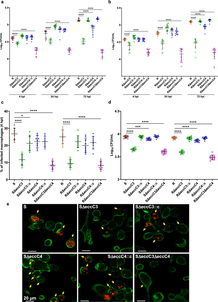
Type VII secretion systems participate in protein export, virulence, conjugation, and metabolic regulation. Five subtypes (ESX-1 to ESX-5) exist, each with specific roles and well-characterized secretion profiles in various mycobacterial species. Mycobacterium abscessus, encodes only ESX-3 and ESX-4. Here, single and double M. abscessus mutants lacking the main ATPases EccC3 and EccC4 were used to define ESX-3 and ESX-4 contributions to substrate secretion and virulence. Our results demonstrate that EsxG/H secretion depends entirely on ESX-3, whereas both ESX-3 and ESX-4 secrete EsxU/T. Furthermore, two newly identified PE/PPE substrates (MAB_0046/MAB_0047) require ESX-3 for secretion. Functional complementation restored secretion and revealed subpolar localization of these systems. Macrophage infections showed that ESX-3 and ESX-4 contribute to bacterial internalization, phagosomal escape, and intracellular survival. In mice, infections with eccC3- and/or eccC4-deletion mutants resulted in complete survival and reduced bacterial loads in the lungs. These findings demonstrate that both ESX systems drive M. abscessus pathogenicity.


Conflict of interest statement

Competing interests: The authors declare no competing interests.

Figures

Fig. 1
Fig. 1. The ESX-3 and ESX-4 secretion systems in M. abscessus.
a Schematic representation of the genetic organization of the esx-3 and esx-4 loci. The genes encoding the different ESX core components are shown using different colors. Genes encoding the secreted substrates are in gray. MAB_3755c (dark gray) encodes a protein with unknown function. b Model of the ESX-3/4 core components using the same color code than in (a). The ATPase domains of EccC3/EccC4 are indicated with triangles.
Fig. 2
Fig. 2. Analysis of secretion proteins in M. abscessus lacking ESX-3 and/or ESX-4.
Immunoblot analysis of bacterial total lysates (Pellet) and secreted proteins (Supernatant) in Mab S (lane 1), ΔeccC3 (lane 2), ΔeccC4 (lane 3), and ΔeccC3/ΔeccC4 (lane 4). Immunoblots illustrating the secretion profile of EsxH (a), PPE4 (b), EsxT (c), EsxH2 (d), MAB_0664 (e), and MAB_0047 (f). Anti-Ag85 antibodies were used as loading controls to verify that the secretion of Ag85 is not affected by the disruption of ESX-3 or ESX-4. GroEL2 serves as a loading control for total lysates and to validate the absence of bacterial lysis. This figure presents data from experimental replicate 1 only. The secreted proteins were tagged with HA tag and expressed from integrative plasmids. Specific proteins were identified using anti-HA antibodies. The design of the fusion proteins and their tags is shown above each blot: (a) EsxG/EsxH-HA, (b) PE5-PPE4-HA-EspG3-STREP, (c) EsxU/EsxT-HA, (d) EsxG2/EsxH2-HA, (e) MAB_0664-HA, (f) MAB_0046-MAB_0047-HA. g Quantification of the bands present in the supernatant fractions shown in panels (a–f) using the Fiji software (mean gray values) (n = 3). h Secretion analysis of the same substrates in complemented strains expressing either EccC3-mNeonGreen or EccC4-mNeonGreen. Data are mean ± SD.
Fig. 3
Fig. 3. Subcellular localization of ESX-3 and ESX-4 in M. abscessus.
Immunofluorescence microscopy of Mab ΔeccC3 and ΔeccC4 mutants producing EccC3 (a) or EccC4 (b) fused to mNeonGreen. Insets represent individual bacilli. Heat maps indicating the localization patterns of fluorescent foci in 50–100 Mab cells are provided. c Detection of EccC3-HA and EccC4-HA in crude lysates of Mab R and S lacking either eccC3eccC3) or eccC4eccC4) carrying the eccC3-HA and eccC4-HA constructs. The genes were under the control of the hsp60 promoter and integrated into the genome. Proteins were detected using Western blot and anti-HA antibodies (upper panel) or anti-KasA antibodies (lower panel, included as a loading control). Positions of EccC3-HA and EccC4-HA are indicated. Lysates of ΔeccC3 and ΔeccC4 mutants complemented with either EccC3-HA or EccC4-HA (d) and EccC3-mNeonGreen or EccC4-mNeonGreen (e) were subjected to sub-cellular fractionation. The fractions were probed with anti-HA antibodies or anti-mNeonGreen antibodies to detect the respective fusion proteins, while anti-GroEL2 antibodies were used as a cytosolic marker. TL, total lysate; CW, cell wall; PM, plasma membrane; Cyt, cytosol. Positions of EccC3-HA or EccC3-mNeonGreen and EccC4-HA or EccC4-mNeonGreen are indicated.
Fig. 4
Fig. 4. EccC3 is crucial for the uptake and intracellular replication of M. abscessus.
a Infection assays with THP-1 macrophages using fluorescent Mab S-derived strains (MOI of 2:1). CFU were determined at 4, 24, and 72 hpi. The data are presented as mean values from three independent experiments (n = 12). Statistical significance was determined using Tukey’s multiple comparisons test: ****, P < 0.0001. b THP-1 cells were infected with the different fluorescent Mab R (MOI of 2:1). CFU counts were similarly determined at 4, 24, and 72 hpi. The data are presented as mean values from three independent experiments (n = 12). Statistical significance was determined using Tukey’s multiple comparisons test: ****, P < 0.0001. c Percentage of infected macrophages was determined for each of the S- or R-derived mutants and their complemented strains. Data represent mean values from three independent experiments, each performed in triplicate (n = 9). Statistical significance was determined using Tukey’s multiple comparisons test: *, P < 0.05, ****, P < 0.0001. d Adhesion assays to evaluate the binding of eccC mutant strains to THP-1 macrophages. Macrophages were pre-cooled to prevent bacterial internalization and exposed to the bacterial strains at a MOI of 100 for 1 hr at 4 °C to enhance bacteria-cell contact. CFUs were quantified to assess adhesion. The data reflect mean values from three independent experiments, each in quadruplicate (n = 12). Statistical analysis was assessed using the one-tailed Mann-Whitney test: ***, P < 0.001, ****, P < 0.0001. e Immunofluorescence microscopy images were taken after 72 hpi, using a confocal microscope at 40x magnification, show macrophages (stained green) infected with various Mab strains (red). Yellow arrows point to mycobacteria-infected cells. Data are mean ± SD.
Fig. 5
Fig. 5. Both ESX-3 an ESX-4 are required for blocking phagosome maturation and phagosomal escape.
a Co-localization of Mab S, SΔeccC3, SΔeccC3::c, SΔeccC4, SΔeccC4::c, SΔeccC3/ΔeccC4, and heat-killed strains (red) with the acidotropic dye LysoTracker (green) in infected THP-1 cells at 20 hpi. Data are presented as means from three independent experiments (n = 9). Statistical significance was assessed using Tukey’s test: ***, P <  0.0001, ****, P < 0.00001. b Acidic compartments were visualized by confocal microscopy using LysoTracker, a fluorescent marker for acidic compartments, in infected macrophages. Co-localization of red fluorescent signals from Mab S, SΔeccC3, SΔeccC3::c, SΔeccC4, SΔeccC4::c, SΔeccC3/ΔeccC4, and heat-killed bacteria with LysoTracker was observed in infected THP-1 cells. Yellow coloring indicated co-localization of red and green labeling. c Percentage of infected macrophages containing at least one positively-stained phagosome (Gal-3+). The values represent the mean of 1000 infected cells analyzed from three different experiments (n = 10). P values were determined by ANOVA with Tukey’s test; ****, P < 0.0001. d IL-1β production during THP-1 infection by Mab S, SΔeccC3, SΔeccC3::c, SΔeccC4, SΔeccC4::c, SΔeccC3/ΔeccC4, and heat-killed strains. The data reflect mean values from three independent experiments, each in quadruplicate (n = 12). P values were determined by ANOVA with Tukey’s test; ****, P < 0.0001. Data are mean ± SD.
Fig. 6
Fig. 6. Effect of ESX-3 and/or ESX-4 depletion on M. abscessus pathogenicity in mice.
C3HeB/FeJ mice were infected intravenously with 2 × 10⁷ CFU/mouse of WT S, SΔeccC3, SΔeccC4, and complemented strains, as well as with SΔeccC3/ΔeccC4. At 1, 7, 14, and 21 dpi. Lungs (a), kidneys (b), spleen (c), and liver (d) were collected for CFU determination. Five mice were used per group (n = 5 mice) (three for day 1, n = 3 mice). Differences between means were analyzed by unpaired t test (a–d) using GraphPad Prism. e Temporal evolution of mice body weights. Numbers in the graph indicate the number of mice analyzed in the experiment (n = 10 mice). f Survival curves of mice infected with WT S, SΔeccC3, SΔeccC4, and SΔeccC3/ΔeccC4 (n = 5 mice per condition). Results in (e) and (f) were generated from two independent experiments. Five mice per group (three for day 1) were used, and differences between means were analyzed by two-way ANOVA with Tukey post-test for multiple comparisons (e). n.s., non-significant; *, P < 0.05; **, P < 0.01; ***, P < 0.001; ****, P < 0.0001. Survival statistics were assessed using the log-rank (Mantel-Cox) test for Kaplan-Meier survival curves (f); ****, P < 0.0001. Data are mean ± SD.
Fig. 7
Fig. 7. Effects of ESX-3 and ESX-4 depletion on M. abscessus pathogenesis.
Mab possesses two T7SSs, ESX-3 and ESX-4. EccC3 and EccC4 are major membrane components of these machineries and are involved in substrate recognition. Following infection, WT bacilli are internalized in macrophage phagosomes and block fusion with acidic compartments. In these non-acidified compartments, ESX-secreted effectors, such as EsxU/EsxT, contribute to phagosomal membrane damage, allowing Mab to escape the phagosome and enter the cytosol. Here, the bacilli trigger the inflammasome, multiply, disseminate, and induce pathology, ultimately leading to the killing of the infected host. In this study, we show that both single and double eccC mutants most likely fail to block the phagolysosomal fusion process. In these mature phagosomes, ESX-3 and ESX-4 mutants cannot withstand the acidic pH and are eliminated by the macrophage, which can be linked to the incapacity to secrete effectors and/or to the inability to maintain metal homeostasis (for ESX-3). In mice, this translates into highly attenuated strains unable to colonize the lungs, thereby prolonging mice survival.

References

    1. Bitter, W. et al. Systematic genetic nomenclature for type VII secretion systems. PLoS Pathog.5, e1000507 (2009). - PMC - PubMed
    1. Pajuelo, D. et al. Toxin secretion and trafficking by Mycobacterium tuberculosis. Nat. Commun.12, 6592 (2021). - PMC - PubMed
    1. Tufariello, J. M. et al. Separable roles for Mycobacterium tuberculosis ESX-3 effectors in iron acquisition and virulence. Proc. Natl. Acad. Sci. USA113, E348–E357 (2016). - PMC - PubMed
    1. Siegrist, M. S. et al. Mycobacterial Esx-3 requires multiple components for iron acquisition. mBio5, e01073–01014 (2014). - PMC - PubMed
    1. Pym, A. S., Brodin, P., Brosch, R., Huerre, M. & Cole, S. T. Loss of RD1 contributed to the attenuation of the live tuberculosis vaccines Mycobacterium bovis BCG and Mycobacterium microti. Mol. Microbiol. 46, 709–717 (2002). - PubMed

MeSH terms

Substances

LinkOut - more resources