. 2025 Mar;26(5):1406-1421.
doi: 10.1038/s44319-025-00383-y. Epub 2025 Feb 3.

Type-2 innate signals are dispensable for skeletal muscle regeneration and pathology linked to Duchenne muscular dystrophy

Affiliations

Type-2 innate signals are dispensable for skeletal muscle regeneration and pathology linked to Duchenne muscular dystrophy

Melina Messing et al. EMBO Rep. 2025 Mar.

Abstract

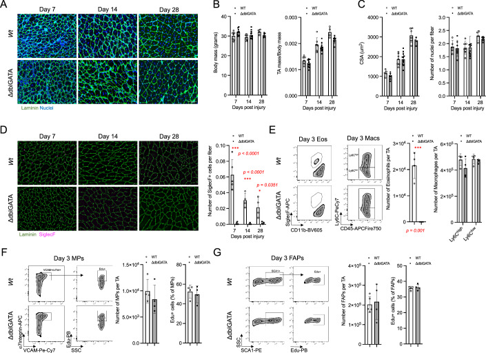
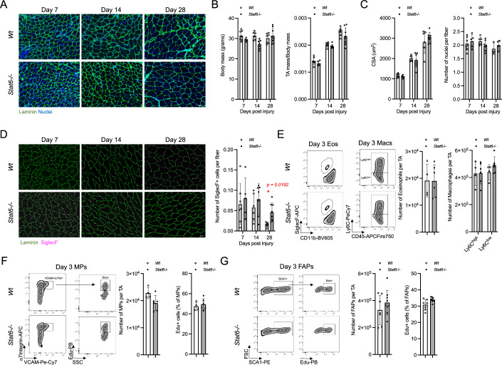
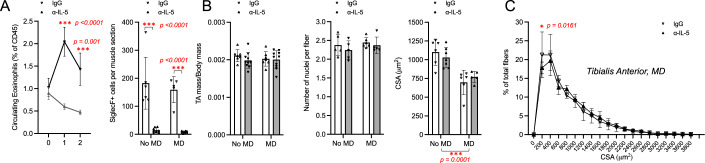
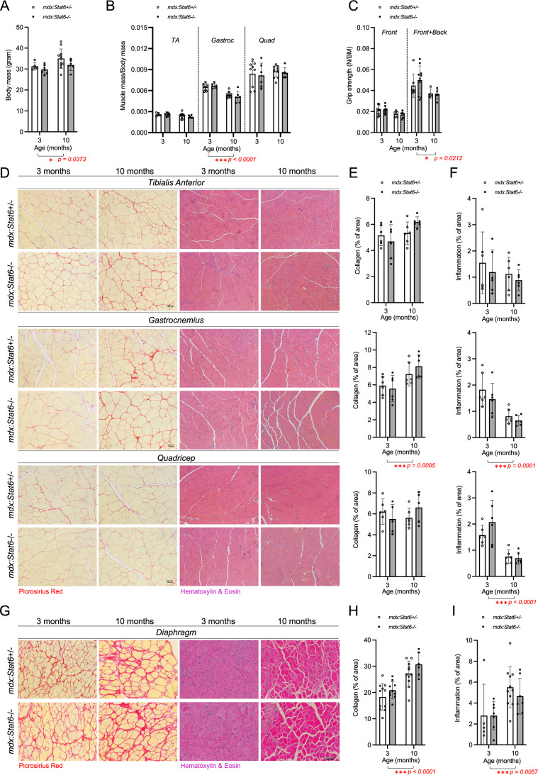
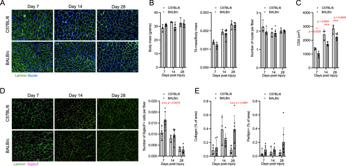
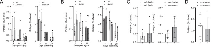
Immune responses play an integral role in skeletal muscle regeneration. In the genetically inherited muscle disease Duchenne muscular dystrophy (DMD), muscle regeneration is disrupted, leading to chronic inflammation, fibrosis, and early mortality. Previously, it has been suggested that type-2 innate immune cells, particularly eosinophils and their production of IL-4, play an essential role in effective muscle regeneration after acute injury. We here re-investigate the role of eosinophils in skeletal muscle repair using mice deficient in eosinophils (ΔdblGATA), or deficient in IL-4R/IL-13R signaling through STAT6 (Stat6-/-). We show that neither deficiency has an impact on skeletal muscle regeneration in response to acute injury as quantified by fiber size, immune cell infiltration, or muscle-resident stem cell proliferation. We also investigate the role of STAT6 signaling in mdx:Stat6-/- mice, a model of DMD and, again, find that ablation of STAT6 signaling has no effect on the rate or severity of fibrotic scar formation or disease progression. In contrast to previous models, our data suggest a negligible role for eosinophils and STAT6 signaling in skeletal muscle regeneration after acute or chronic injury.

Keywords: Duchenne Muscular Dystrophy (DMD); Eosinophils; STAT6 Signaling; Skeletal Muscle Regeneration.


Conflict of interest statement

Disclosure and competing interests statement. The authors declare no competing interests.

Figures

Figure 1
Figure 1. Eosinophils are dispensable for muscle regeneration.
Tibialis anterior (TA) muscles of wild-type (Wt) or ΔdblGATA mice were injured with barium chloride (BaCl2) and harvested to assess muscle regeneration at 7-, 14-, and 28-days post injury (AD). Representative histological laminin and nuclei (DAPI) staining of TA sections (A). Mouse body mass and TA mass (normalized to body mass) measurements (B). Quantification of cross-sectional area (CSA) and number of nuclei per fiber (C). Representative histological SiglecF staining and quantification of SiglecF+ cells in TA muscle sections (D). Flow cytometry representative gating and cell number quantification of eosinophils (gated as live/CD45+/CD11blow/SiglecF+) and macrophages (gated on live/CD45+/Lin-/Ly6Chigh/low) in TA muscles at 3 days post-BaCl2 injury (E). Flow cytometry representative gating, cell number and % proliferation (% EdU+) quantification of myogenic progenitors (MPs) (gated on live/CD45-/CD31-/SCA-1-/ɑ7 integrin+/VCAM+) (F). Flow cytometry representative gating, cell number and % proliferation (% EdU+) quantification of fibro/adipogenic progenitors (FAPs) (gated on live/CD45-/CD31-/SCA-1+) (G). Number of mice (biological replicates) per group: n = 4–9. Two independent replicates were performed for each experiment. *p < 0.05; ***p < 0.001 by two-tailed, two-sample unequal variance Student’s t-test or two-way ANOVA with post hoc testing (Šidak multiple comparison). Scale bars show 100 µm. Error bars represent SD. Source data are available online for this figure.
Figure 2
Figure 2. STAT6 signaling is dispensable for muscle regeneration.
Tibialis anterior (TA) muscles of wild-type (Wt) or Stat6−/− mice were injured with barium chloride (BaCl2) and harvested to assess muscle regeneration at 7-, 14-, and 28-days post injury (AD). Representative histological laminin and nuclei (DAPI) staining of TA sections. Mouse body mass and TA mass (normalized to body mass) measurements (B). Quantification of cross-sectional area (CSA) and number of nuclei per fiber (C). Representative histological SiglecF staining and quantification of SiglecF+ cells in TA muscle sections (D). Flow cytometry representative gating and cell number quantification of eosinophils (gated as live/CD45+/CD11blow/SiglecF+) and macrophages (gated on live/CD45+/Lin-/Ly6Chigh/low) in TA muscles at 3 days post-BaCl2 injury (E). Flow cytometry representative gating, cell number and % proliferation (% EdU+) quantification of myogenic progenitors (MPs) (gated on live/CD45-/CD31-/SCA-1-/ɑ7 integrin+/VCAM+) (F). Flow cytometry representative gating, cell number and % proliferation (% EdU+) quantification of fibro/adipogenic progenitors (FAPs) (gated on live/CD45-/CD31-/SCA-1+) (G). Number of mice (biological replicates) per group: n = 4–8. Two independent replicates were performed for each experiment. *p < 0.05 by two-way ANOVA with post hoc testing (Šidak multiple comparison). Scale bars show 100 µm. Error bars represent SD. Source data are available online for this figure.
Figure 3
Figure 3. STAT6 signaling and eosinophils are dispensable for the repair of chronic muscle injury.
Flow cytometry cell number quantification of eosinophils (gated as live/CD45+/CD11blow/SiglecF+) and macrophages (gated on live/CD45+/Lin-/Ly6Chigh/low), cell number and % proliferation (% EdU+) quantification of myogenic progenitors (MPs) (gated on live/CD45-/CD31-/SCA-1-/ɑ7 integrin+/VCAM+), and cell number quantification of fibro/adipogenic progenitors (FAPs) (gated on live/CD45-/CD31-/SCA-1+) in pooled undamaged tibialis anterior TA, gastrocnemius (Gastroc) and quadricep (Quad) muscles (A). At age 3 months, the right TA of mdx:Stat6+/− and mdx:Stat6−/− mice were micro-damaged (MD) while the left TA was not damaged (No MD); Tissues were collected at 14 weeks of age (BE). Representative histological laminin and SiglecF staining and quantification of SiglecF+ cells in TA muscle sections (B). Representative histological laminin and nuclei (DAPI) staining of TA muscle sections (C). TA mass (normalized to body mass) measurements, quantification of number of nuclei per fiber, cross-sectional area (CSA) and binned CSA (D, E). Number of mice (biological replicates) per group: n = 4–7. Two independent replicates were performed for each experiment. *p < 0.05; **p < 0.001 by two-way ANOVA with post hoc testing (Šidak multiple comparison). Scale bar represents 100 µm. Error bars represent SD. Source data are available online for this figure.
Figure 4
Figure 4. Eosinophils are dispensable for the repair of chronic muscle injury.
Quantification of circulating eosinophil frequency and SiglecF+ cells per MD and No MD TA sections of ɑIL-5 or IgG intraperitoneally injected mdx mice (A). TA mass (normalized to body mass) measurements, quantification of number of nuclei per fiber, cross-sectional area (CSA) and binned CSA (B, C). Number of mice (biological replicates) per group: n = 4–7. Two independent replicates were performed for each experiment. *p < 0.05; ***p < 0.001 by two-way ANOVA with post hoc testing (Šidak multiple comparison). Scale bar represents 100 µm. Error bars represent SD. Source data are available online for this figure.
Figure 5
Figure 5. STAT6 signaling does not contribute to DMD pathology.
At age 3 months and 10 months, mouse body mass (A), muscle mass (normalized to body mass) of tibialis anterior (TA), gastrocnemius (Gastroc) and quadricep (Quad), and grip strength force (normalized to body mass) were measured in mdx:Stat6+/− and mdx:Stat6−/− mice (C). Representative histological picrosirius red (PSR) and hematoxylin and eosin (H&E) staining of TA (top), Gastroc (middle), and Quad (bottom) at 3 months and 10 months (D). Quantification of % collagen based on PSR staining (E) and inflammation based on H&E staining (F) of TA, Gastroc and Quad at age 3 months and 10 months. Representative histological PSR and H&E of diaphragm (G) and % collagen and inflammation quantification (H, I). Number of mice (biological replicates) per group: n = 6–11. Two independent replicates were performed for each experiment. *p < 0.05; ***p < 0.001 by two-way ANOVA with post hoc testing (Šidak multiple comparison). Scale bar represents 100 µm. Error bars represent SD. Source data are available online for this figure.
Figure 6
Figure 6. Muscle regeneration differs in BALB/c and C57BL/6 mice.
Tibialis anterior (TA) muscles of C57BL/6 and BALB/c mice were injured with barium chloride (BaCl2) and body mass, TA mass (normalized to body mass), number of nuclei per fiber, and cross-sectional area (CSA) were quantified to assess muscle regeneration at 7-, 14-, and 28-days post injury (AC). To assess eosinophil infiltration, collagen and adipocyte deposition, number of SiglecF+ cells, collagen (%of area), and perilipin (% of area) were quantified respectively (D, E). Number of mice per group: n = 3–7. Two independent replicates were performed for each experiment. *p < 0.05; ***p < 0.001 by two-way ANOVA with post hoc testing (Šidak multiple comparison). Error bars represent SD.
Figure EV1
Figure EV1. Adipocyte and collagen deposition analysis.
Quantification of % perilipin and % collagen in TA muscle sections Tibialis anterior (TA) muscles of wild-type (Wt) or ΔdblGATA mice at 7-, 14-, and 28-days post injury after muscle injury with barium chloride (BaCl2) (A). Quantification of % perilipin and % collagen in TA muscle sections muscles of wild-type (Wt) or Stat6−/− mice at 7-, 14-, and 28-days post injury after muscle injury with barium chloride (BaCl2) (B). At age 3 months, the right TA of mdx:Stat6+/− and mdx:Stat6−/− mice were micro-damaged (MD).Tissues were collected at 14 weeks of age. Quantification of % perilipin and % collagen in MD TA muscle sections (C). Quantification of % perilipin in 10 months old mdx:Stat6+/− and mdx:Stat6−/− mice (D). Number of mice per group: n = 3–4. Error bars represent SD.
Figure EV2
Figure EV2. Analysis of intermediate loss of STAT6.
Tibialis anterior (TA) muscles of wild-type (Wt), Stat6+/− or Stat6−/− mice were injured with barium chloride (BaCl2) and body mass, TA mass (normalized to body mass), cross-sectional area (CSA), number of nuclei per fiber and number of SiglecF+ cells was quantified to assess muscle regeneration at 7-, 14-, and 28-days post injury (A). At age 3 months, mdx, mdx:Stat6+/− and mdx:Stat6−/− mice were compared in terms of body mass, TA/body mass, CSA and nuclei per fiber as well as % collagen deposition in the diaphragm (B). Number of mice per group: n = 3–7. Error bars represent SD.
Figure EV3
Figure EV3. Representative flow cytometry gating strategies.
Muscle resident cells gating strategy (A). Eosinophil and macrophage gating strategy (B). FVD fixable viability dye, Lin lineage.

References

    1. Akimoto T, Numata F, Tamura M, Takata Y, Higashida N, Takashi T, Takeda K, Akira S (1998) Abrogation of bronchial eosinophilic inflammation and airway hyperreactivity in signal transducers and activators of transcription (STAT)6-deficient mice. J Exp Med 187:1537–1542 - PMC - PubMed
    1. Annunziato F, Romagnani C, Romagnani S (2015) The 3 major types of innate and adaptive cell-mediated effector immunity. J Allergy Clin Immunol 135:626–635 - PubMed
    1. Arnold L, Henry A, Poron F, Baba-Amer Y, Van Rooijen N, Plonquet A, Gherardi RK, Chazaud B (2007) Inflammatory monocytes recruited after skeletal muscle injury switch into anti-inflammatory macrophages to support myogenesis. J Exp Med 204:1057–1069 - PMC - PubMed
    1. Babaeijandaghi F, Cheng R, Kajabadi N, Soliman H, Chang CK, Smandych J, Tung LW, Long R, Ghassemi A, Rossi FM (2022) Metabolic reprogramming of skeletal muscle by resident macrophages points to CSF1R inhibitors as muscular dystrophy therapeutics. Sci Transl Med 14:eabg7504 - PubMed
    1. Babaeijandaghi F, Kajabadi N, Long R, Tung LW, Cheung CW, Ritso M, Chang CK, Cheng R, Huang T, Groppa E et al (2023) DPPIV+ fibro-adipogenic progenitors form the niche of adult skeletal muscle self-renewing resident macrophages. Nat Commun 14:8273 - PMC - PubMed

MeSH terms

Substances

LinkOut - more resources