Review
. 2025 Feb 3;16(1):1307.
doi: 10.1038/s41467-024-55016-0.

Reward signals in the motor cortex: from biology to neurotechnology

Affiliations
Review

Reward signals in the motor cortex: from biology to neurotechnology

Gerard Derosiere et al. Nat Commun. .

Abstract

Over the past decade, research has shown that the primary motor cortex (M1), the brain's main output for movement, also responds to rewards. These reward signals may shape motor output in its final stages, influencing movement invigoration and motor learning. In this Perspective, we highlight the functional roles of M1 reward signals and propose how they could guide advances in neurotechnologies for movement restoration, specifically brain-computer interfaces and non-invasive brain stimulation. Understanding M1 reward signals may open new avenues for enhancing motor control and rehabilitation.


Conflict of interest statement

Competing interests: The authors declare no competing interests.

Figures
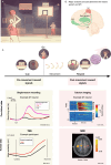
Fig. 1
Fig. 1. The influence of reward on motor behavior and M1 activity.
A Influence of reward on motor behavior. Consider a basketball player about to make a potentially game-winning shot. The player glances at the gleaming trophy before making the throw. Upon the referee’s whistle, the ball is thrown and successfully lands in the basket, leading to a victory and the team receiving the trophy. This scenario exemplifies how motor behaviors are directed and shaped by rewards. The context of the reward (e.g., making a shot to win the trophy in the Olympics final) directly influences the kinematics of the movements performed. Furthermore, the outcomes of these movements (e.g., a successful or failed shot) significantly affect future adjustments in motor commands, thereby influencing learning. B Anatomical routes between reward system structures and M1. Several key brain structures, consistently associated with reward processing, form the “reward system” (highlighted in green). These include the midbrain’s dopaminergic structures, particularly the ventral tegmental area (VTA), the basal ganglia (especially the ventral striatum), and the orbitofrontal cortex (OFC, with its medial part depicted here, also known as the ventromedial prefrontal cortex or vmPFC). Crucially, these structures connect to M1 (marked in red) through several bi-directional circuits, providing essential anatomical pathways for bidirectional influences between the reward system and M1. The brain image in this panel was extracted from BioRender.com and released under a Creative Commons Attribution-NonCommercial-NoDerivs 4.0 International license (Raffin, E. (2024) https://BioRender.com/v49b404). C Pre-movement and post-movement reward signals in M1. The time course at the top outlines the key stages of a reward-based task. Initially, motivational cues typically signal the reward at stake in a specific context. In the basketball scenario, this could be the gleaming trophy. A go cue then signals the need to execute the movement; this corresponds to the referee’s whistle in the basketball example. Once the movement is executed (i.e., throwing the ball to the basket), a reward is given if the outcome is successful, reinforcing the motor behavior. In our example, the rewards would be an increased score, the cheering of the crowd, and ultimately, winning the trophy. Neuroscientists typically categorize reward signals into two broad types based on whether they occur before or after throwing the ball in our example. Signals that arise before movements are often associated with an activational or motivational role of reward. In the basketball scenario, these signals would reflect the expected consequences of the shoot given the context. Signals occurring after movements are linked to reinforcement learning. In the basketball example, these signals would drive adaptive processes allowing movement correction given the outcome of the shoot. Numerous studies now confirm that M1 exhibits both pre- and post-movement reward signals. Specifically, pre-movement M1 activity scales with reward magnitude (i.e., from small to large rewards); this has been demonstrated in both pre-clinical, single-neuron studies in macaques (top left graph) and in human studies using transcranial magnetic stimulation to probe motor excitability (bottom left graph). In addition, post-movement M1 activity is modulated by the outcome of the movement both at the single-neuron level in mice (top right graph) and at the population level using fMRI (bottom right graph, note also the concurrent premotor cortex activation). The neuron depicted in the top-right graph is provided as an example, but note that other neurons in M1 exhibit opposite changes (i.e., modulation following failure and not success,–. Images from previous studies are adapted with permission.
Fig. 2
Fig. 2. Application of Pre- and Post-Movement Reward Signals in M1 to Enhance BCIs.
a BCI scenario. The example shows a BCI scenario in which a user with arm paralysis controls a robotic arm through brain activity (inspired by the work of ref. ). b Neural signals. The neural signals in this example are single-unit recording, though similar approaches have also been demonstrated using local field potentials (LFPs). c Multi-stage decoder. (i) The neural data is initially processed to decode the context of the reaching movement—whether it occurs in a rewarded or non-rewarded setting. (ii) Depending on the context, one of two distinct decoders is used. In this example, the user is in a rewarded context (e.g., attempting to drink from a fresh bottle on a hot day). (iii) If the trial fails (e.g., the bottle is dropped, and the user cannot drink), the lack of a reward is detected by an outcome decoder. This information is then used to update the context-specific decoder chosen in the previous step, allowing the BCI to adapt and improve over time. Representation of the BCI was adapted from.
Fig. 3
Fig. 3. Translational roadmap toward reward state-dependent stimulation of M1.
The top row represents 3 key properties of M1 reward signals: timing-selectivity, outcome-dependence, and functional heterogeneity of reward encoding. Note that the plots are provided for illustration purposes of the concepts based on previous literature but do not reflect actual data. These neuroscientific observations naturally lead to a series of expected features for NIBS technologies aiming at delivering reward-state dependent M1 stimulation. More precisely, we suggest that the employed technology will need to enable rapid triggering, flexible adjustment of parameters according to behavior, and subthreshold, low-intensity stimulation. In the bottom row, we highlight what we consider as the key options to satisfy each constraint and propose an integrated solution based on this analysis, with the ultimate goal of achieving non-invasive and reward-state dependent stimulation of M1. Brains in the right panels were extracted from BioRender.com and released under a Creative Commons Attribution-NonCommercial-NoDerivs 4.0 International license (Raffin, E. (2024) https://BioRender.com/v49b404).

Similar articles

Cited by

References

    1. Manohar, S. G. et al. Reward pays the cost of noise reduction in motor and cognitive control article reward pays the cost of noise reduction in motor and cognitive control. Curr. Biol.25, 1707–1716 (2015). - PMC - PubMed
    1. Codol, O., Holland, P. J., Manohar, S. G. & Galea, J. M. Reward-based improvements in motor control are driven by multiple error-reducing mechanisms. J. Neurosci.40, 3604–3620 (2020). - PMC - PubMed
    1. Neiman, T. & Loewenstein, Y. Reinforcement learning in professional basketball players. Nat. Commun.2, 569 (2011). - PMC - PubMed
    1. Wu, H. G., Miyamoto, Y. R., Castro, L. N. G., Ölveczky, B. P. & Smith, M. A. Temporal structure of motor variability is dynamically regulated and predicts motor learning ability. Nat. Neurosci.17, 312–321 (2014). - PMC - PubMed
    1. Dhawale, A. K., Miyamoto, Y. R., Smith, M. A. & Ölveczky, B. P. Adaptive regulation of motor variability. Curr. Biol.29, 3551–3562 (2019). - PMC - PubMed

LinkOut - more resources