. 2025 Feb 3;16(1):63.
doi: 10.1038/s41419-025-07375-9.

FAM210B activates STAT1/IRF9/IFIT3 axis by upregulating IFN-α/β expression to impede the progression of lung adenocarcinoma

Affiliations

FAM210B activates STAT1/IRF9/IFIT3 axis by upregulating IFN-α/β expression to impede the progression of lung adenocarcinoma

Xuejuan Gao et al. Cell Death Dis. .

Abstract

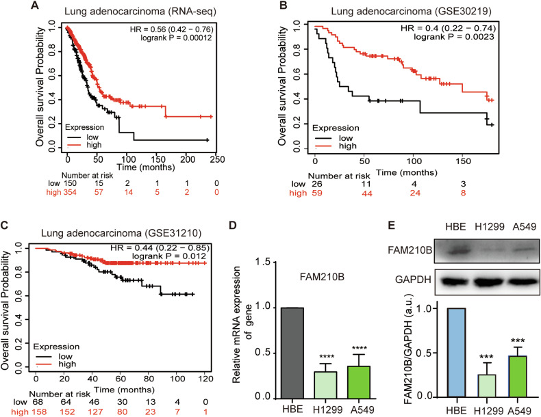
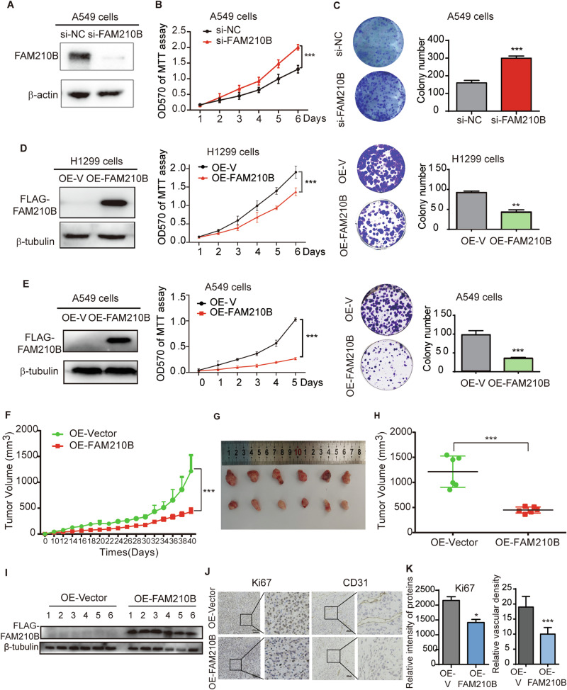
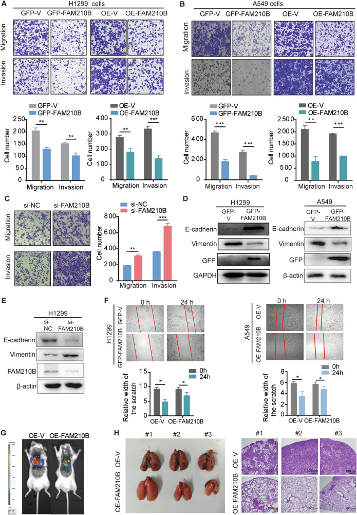
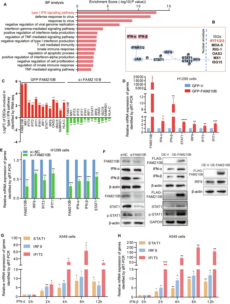
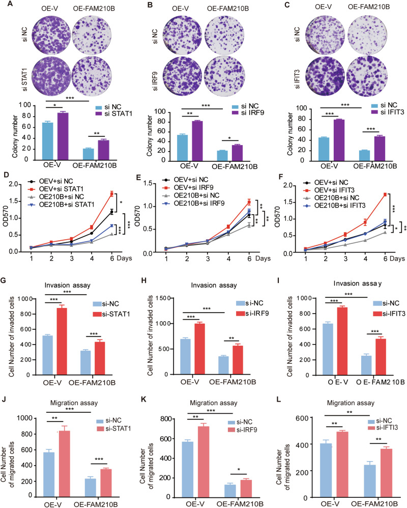
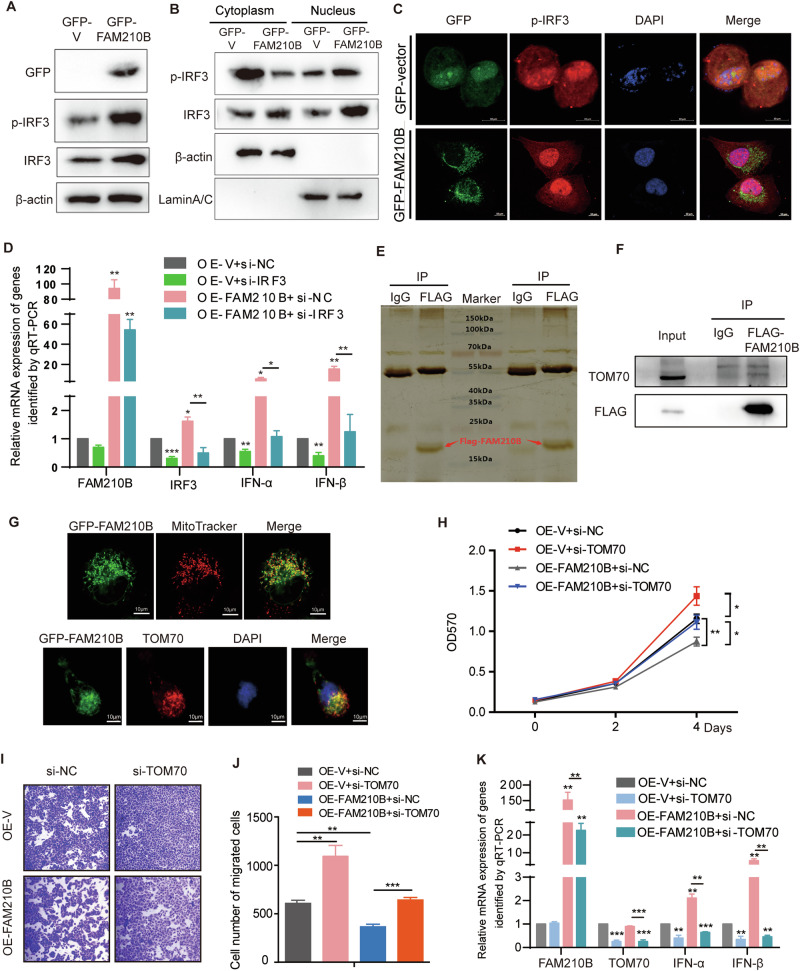
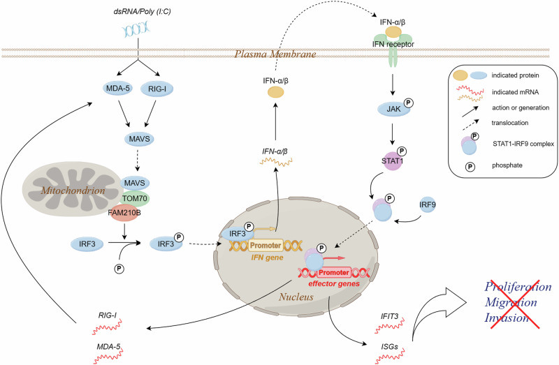
FAM210B (family with sequence similarity 210 member B) is a novel protein that has been linked to tumor development. However, its role and underlying mechanisms in lung adenocarcinoma (LUAD) progression remain largely unexplored. In this study, FAM210B was observed to be down-regulated in LUAD cells. Analyses of public datasets revealed that decreased expression of FAM210B predicts poor survival. Accordingly, in vitro and in vivo studies have confirmed the inhibitory role of FAM210B on the growth and tumor metastasis of LUAD cells. RNA-seq analysis further indicated that FAM210B plays a role in regulating innate immune-related signaling pathways in LUAD cells, particularly involving the production of type I interferon (IFN-α/β). Specifically, FAM210B activates STAT1/IRF9/IFIT3 axis by upregulating IFN-α/β expression, leading to the inhibition of proliferation and migration of LUAD cells. Furthermore, TOM70 (Translocase of outer mitochondrial membrane 70, also named as TOMM70) has been identified as a functional interacting partner of FAM210B in its modulation on the expression of IFN-α/β, as well as the proliferative and metastatic phenotypes of LUAD cells. In conclusion, our study indicates that FAM210B is an important suppressor of cellular viability and mobility during lung cancer progression.


Conflict of interest statement

Competing interests: The authors declare no competing interests.

Figures

Fig. 1
Fig. 1. FAM210B expression level was positively correlated with overall survival in LUAD patients.
Kaplan-Meier survival analysis of the relationship between FAM210B expression and overall survival in TCGA-LUAD RNA-seq data (A), GSE30219 (B), and GSE31210 (C), with high expression of FAM210B denoted in red and low expression of FAM210B in black. D qRT-PCR was used to analyze the expression levels of FAM210B gene in HBE, H1299, and A549 cell lines. E Western blot analysis of FAM210B protein expression in HBE, H1299, and A549 cell lines (Upper). Quantitative analysis of FAM210B protein expression of three independent repeated experiments (Lower). Data of D-E were represented as mean ± SEM of three independent experiments. Statistical analysis was performed using Student’s t-test. ***P < 0.001, ****P < 0.0001.
Fig. 2
Fig. 2. FAM210B suppressed LUAD cell proliferation in vitro and LUAD tumor growth in vivo.
A Western blot analysis of FAM210B protein expression after its knockdown in A549 cells. B MTT assay of A549 cells after FAM210B knockdown. C Colony formation assays of A549 cells after FAM210B knockdown. D H1299 cells stably overexpressing FAM210B (OE-FAM210B, left panel) were subjected to MTT (middle panel) and colony formation assays (right panel). E A549 cells stably overexpressing FAM210B (OE-FAM210B, left panel) were subjected to MTT (middle panel) and colony formation assays (right panel). FH FAM210B inhibited LUAD tumor growth in vivo. Assays were performed with the injection of either OE-FAM210B A549 cells or the control OE-Vector A549 cells. The results presented for the indicated nude mouse groups are: evolution of tumor volume measured at different time points (F, 40 days after subcutaneous injection of A549 OE-V and OE-FAM210B cells, the tumor volume neared the upper limit allowed by animal ethics guidelines, prompting the termination of the experiment), images of tumors at the endpoints of time (G), and the statistical representation of tumor volumes illustrated in (G, H). I Western blot assays were performed on the tumor tissues from the indicated nude mouse groups to confirm the overexpression of FAM210B. J IHC analyses of Ki67 and CD31 were performed on tumor tissues from each group, and representative images were presented. K Quantitative analysis of Ki67 intensity in (J) and quantitative analysis of vascular density based on CD31 staining in J were performed. Data of AE were represented as mean ± SEM from three independent experiments. Statistical analysis was performed using two-way ANOVA (B, MTT data of D and E, F) and Student’s t-test (C, colony assay of D and E, H, K). *P < 0.05, **P < 0.01, ***P < 0.001.
Fig. 3
Fig. 3. FAM210B suppressed the migration and invasion of LUAD cells in vitro and the tumor metastasis of LUAD tumor in vivo.
A, B Transwell assays were conducted to analyze the migration and invasion abilities of H1299 cells or A549 cells transiently (left) or stably (right) overexpressing FAM210B (upper). The quantitative analysis of invasion and migration in H1299 and A549 cells was shown in the lower panels. **P < 0.01, ***P < 0.001 (Student’s t-test). C Transwell assays were performed in FAM210B knocked-down H1299 cells (left). The quantitative analysis of invasion and migration in left panel was shown in the right panel. **P < 0.01, ***P < 0.001 (Student’s t-test). D, E Western blot was performed on H1299 and A549 cells transiently transfected with GFP-FAM210B or FAM210B siRNA to analyze the expression of EMT markers. F Wound healing experiments were conducted on H1299 cells transiently overexpressing GFP-FAM210B and on A549 cells stably overexpressing FAM210B. The quantitative analysis of the scratch width was presented below the corresponding images (mean ± SEM, n = 3). *P < 0.05 (Student’s t-test). G Representative bioluminescent images of NCG mice acquired 40 days after tail intravenous injection of A549 cells expressing OE-V-Luc or OE-FAM210B-Luc. H Pictures of lungs taken from the indicated groups of nude mice (left), and histological examination of lung tissues using HE staining (right). Scale bar: 100 μm.
Fig. 4
Fig. 4. FAM210B activated the IFN-α/β mediated STAT1/IRF9/IFIT3 signal pathway.
A GO Biological Process (BP) analysis of 113 DEGs regulated by FAM210B, with the top 15 BPs listed. B A schematic representation of the type I IFN signaling pathway. The circled P represents phosphate. C Log (fold change) of mRNA expression level of DEGs involved in the type I IFN signaling pathway, as assayed by RNA-seq with FAM210B overexpressing or knockdown H1299 cells. All the selected genes have a P value of less than 0.01 (**P < 0.01). qRT-PCR assays were used to test the expression levels of several representative members of the type I IFN signaling pathway, along with IFN-α and IFN-β, in H1299 cells overexpressing GFP-FAM210B (D) or following FAM210B knockdown (E) (mean ± SEM, n = 3). F Western blotting was conducted in H1299 cells with FAM210B knockdown or overexpression (OE-FAM210B) to evaluate changes in representative members of the type I IFN signaling pathway. GH qRT-PCR assays were employed to assess the expression levels of STAT1, IRF9, and IFIT3 in LUAD cells under treatment with IFN-α or IFN-β at different time points (n = 2), respectively. Statistical analysis was performed using Student’s t-test. *P < 0.05, **P < 0.01, ***P < 0.001, ****P < 0.0001.
Fig. 5
Fig. 5. STAT1/IRF9/IFIT3 axis was involved in the inhibitory function of FAM210B in the proliferation and migration of LUAD cells.
Colony formation assay of OE-V and OE-FAM210B H1299 cells transfected with si-STAT1 (A), si-IRF9 (B), or si-IFIT3 (C) (top panels). Quantitative analysis of these colony formation of three biological replicates was shown in bottom panels. MTT assays of OE-V and OE-FAM210B H1299 cells transfected with si-STAT1 (D), si-IRF9 (E), or si-IFIT3 (F). Quantitative analysis of transwell assays for invasion and migration in OE-V and OE-FAM210B H1299 cells transfected with si-STAT1(G, J), si-IRF9 (H, K), or si-IFIT3 (I, L) as indicated. All data were represented as mean ± SEM from three independent experiments. Statistical analysis was performed using two-way ANOVA (D, E, F) and Student’s t-test (AC, GL). *P < 0.05, **P < 0.01, ***P < 0.001.
Fig. 6
Fig. 6. FAM210B promoted the expression of IFN-α/β via activating IRF3 and associating with TOM70.
A Western blot assay of the expression of p-IRF3 and IRF3 in H1299 cells transfected with GFP-V or GFP-FAM210B. B Nuclear-cytoplasmic separation assays were used to analyze the expression changes of p-IRF3 in both nuclear and cytoplasmic fractions in H1299 cells transfected with GFP-V or GFP-FAM210B. C IF assay of localization of p-IRF3 in H1299 cells transfected with GFP-V or GFP-FAM210B. Scale bar: 10 μm. D qRT-PCR assays of the indicated genes in OE-V and OE-FAM210B H1299 cells combined with si-IRF3 as indicated. E Silver staining of the immunocomplex immunoprecipitated by FLAG antibody and non-immune IgG antibody. F The interaction of FLAG-FAM210B with TOM70 confirmed by co-IP assay in H1299 OE-FAM210B cells. G Imaging assay was used to assess the colocalization of GFP-FAM210B with mitochondria (MitoTracker Red) in live H1299 cells (upper), and IF assay was performed to detect the colocalization of GFP-FAM210B with TOM70 in H1299 cells transfected with GFP-FAM210B (below). Scale bar: 10 μm. H MTT assay of the cellular viability of OE-V and OE-FAM210B H1299 cells transfected with si-TOM70. I Transwell assays of the migration of OE-V and OE-FAM210B H1299 cells transfected with si-TOM70. The corresponding quantitative analysis of this transwell assay is shown in (J). K qRT-PCR assay of the expression of IFN-α/β in OE-V and OE-FAM210B H1299 cells transfected with si-TOM70. All quantitative analyses are presented as mean ± SEM from three independent experiments. Statistical analysis was conducted using Student’s t-test (D, J, K) and two-way ANOVA (H). *P < 0.05, **P < 0.01, ***P < 0.001.
Fig. 7
Fig. 7. FAM210B inhibits the proliferation and metastasis of lung adenocarcinoma via interacting with TOM70 and promoting IFN-α/β production.
When FAM210B is overexpressed in LUAD cells, it interacts with TOM70, leading to the phosphorylation and nuclear translocation of IRF3. This activation of IRF3 enhances the expression and secretion of IFN-α/β, which then binds to its receptor and activates the STAT1/IRF9 pathway. As a result, the expression of ISGs like IFIT3, MDA-5, and RIG-I is induced, which in turn suppresses LUAD cell proliferation, migration, and invasion. Elevated levels of MDA-5 and RIG-I further stimulate the innate immune response, with FAM210B playing a role in amplifying IFN-α/β expression.

Similar articles

Cited by

References

    1. Lai XN, Li J, Tang LB, Chen WT, Zhang L, Xiong LX. MiRNAs and LncRNAs: dual roles in TGF-beta signaling-regulated metastasis in lung cancer. Int J Mol Sci. 2020;21:1193. - PMC - PubMed
    1. Thai AA, Solomon BJ, Sequist LV, Gainor JF, Heist RS. Lung cancer. Lancet. 2021;398:535–54. - PubMed
    1. Yuan S, Yu Z, Liu Q, Zhang M, Xiang Y, Wu N, et al. GPC5, a novel epigenetically silenced tumor suppressor, inhibits tumor growth by suppressing Wnt/beta-catenin signaling in lung adenocarcinoma. Oncogene. 2016;35:6120–31. - PubMed
    1. Uze G, Schreiber G, Piehler J, Pellegrini S. The receptor of the type I interferon family. Curr Top Microbiol Immunol. 2007;316:71–95. - PubMed
    1. Xue C, Yao Q, Gu X, Shi Q, Yuan X, Chu Q, et al. Evolving cognition of the JAK-STAT signaling pathway: autoimmune disorders and cancer. Signal Transduct Target Ther. 2023;8:204. - PMC - PubMed

MeSH terms

Substances