. 2025 Feb 3;16(1):65.
doi: 10.1038/s41419-025-07394-6.

LDHA-mediated glycolysis in stria vascularis endothelial cells regulates macrophages function through CX3CL1-CX3CR1 pathway in noise-induced oxidative stress

Affiliations

LDHA-mediated glycolysis in stria vascularis endothelial cells regulates macrophages function through CX3CL1-CX3CR1 pathway in noise-induced oxidative stress

Ying Yi et al. Cell Death Dis. .

Abstract

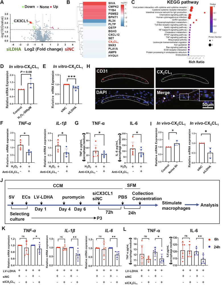
According to the World Health Organization, more than 12% of the world's population suffers from noise-induced hearing loss (NIHL). Oxidative stress-mediated damage to the stria vascularis (SV) is one of the pathogenic mechanisms of NIHL. Recent studies indicate that glycolysis plays a critical role in endothelial cells (ECs)-related diseases. However, the specific role of glycolysis in dysfunction of SV-ECs remain largely unknown. In this study, we investigated the effects of glycolysis on SV-ECs in vitro and on the SV in vivo. Our previous research identified the glycolysis pathway as a potential mechanism underlying the SV-ECs injuries induced by oxidative stress. We further examined the expression levels of glycolytic genes in SV-ECs under H2O2 stimulation and in noise-exposed mice. We found that the gene and protein expression levels of glycolytic-related enzyme LDHA significantly decreased at early phase after oxidative stress injury both in vitro and in vivo, and exhibited anti-inflammatory effects on macrophages (Mφ). Moreover, we analyzed the differential secretomes of SV-ECs with and without inhibition of LDHA using LC-MS/MS technology, identifying CX3CL1 as a candidate mediator for cellular communication between SV-ECs and Mφ. We found that CX3CL1 secretion from SV-ECs was decreased following LDHA inhibition and exhibited anti-inflammatory effects on Mφ via the CX3CR1 pathway. Similarly, the pro-inflammatory effect of LDHA-overexpressing SV-ECs was attenuated following inhibition of CX3CL1. In conclusion, our study revealed that glycolysis-related LDHA was reduced in oxidative stress-induced SV-ECs, and that LDHA inhibition in SV-ECs elicited anti-inflammatory effects on Mφ, at least partially through the CX3CL1-CX3CR1 pathway. These findings suggest that LDHA represent a novel therapeutic strategy for the treatment of NIHL.


Conflict of interest statement

Competing interests: The authors declare no competing interests. Ethics approval: All animal procedures were approved by the Institutional Animal Care and Use Committee of The First Affiliated Hospital, Sun Yat-sen University (SYSU-IACUC-2022-000518), and were conducted in compliance with the National Institutes of Health guidelines for the care and use of laboratory animals. All methods were performed in accordance with the relevant guidelines and regulations.

Figures

Fig. 1
Fig. 1. The function of SV-ECs injury occurred in the acute phase following oxidative stress.
A H&E staining of cochlear lateral walls and quantifications of cross-section area of SV in all cochlear turns by Image J. Significant increases in middle and basal turns of SV after noise exposure were found (n = 7) (Scale bar, 50 μm). B Representative confocal figures of BS-IB4 (green) indicating stronger fluorescence in damaged SV-ECs in 3 days after noise. (Scale bar, 50μm). C Representative confocal pictures of 3-NT (green) showing stronger fluorescence of SV in 3 days post-noise exposure (Scale bar, 50 μm). D Relative mRNA levels of anti-oxidative genes, including SOD1, SOD2 and GSR, were decreased in SVs of cochleae after noise exposure (n = 3). E Angioplasty experiments revealed significant reductions in the branches length, number of junctions, number of nodes, number of meshes and area of endothelial cell angioplasty after stimulation with 100 μM and 500 μM H2O2 for 2 h (scale bar, 500 μm) (n = 3). F Representative images and quantification of transwell migration assays of SV-ECs (scale bar, 200 μm) (n = 5). G Transepithelial electrical resistance (TEER) decreased in SV-ECs treated with 500 μM H2O2 for 2 h (n = 3). H ROS fluorescence intensity was increased in SV-ECs stimulated by 500 μM H2O2 for 2 h and 24 h (n = 4). I Relative mRNA levels of anti-oxidative, including SOD1, SOD2, GSR, CAT and GPX, were decreased in SV-ECs at 2 h following treatment with 500 μM H2O2 (n = 3). The data is expressed as Mean ± SD. *P < 0.05, **P < 0.01, *** P < 0.001, **** P < 0.0001 analyzed by Unpaired t test, SV stria vascularis, ECs endothelial cells, ns not significant, SOD1 Superoxide Dismutase 1, SOD2 Superoxide Dismutase 2, GSR Glutathione-disulfide reductase, GPx Glutathione peroxidases, CAT Catalase, qRT-PCR Quantitative Reverse Transcription Polymerase Chain Reaction.
Fig. 2
Fig. 2. Effects of oxidative stress on glycolysis pathway.
A Schematic diagram illustrating the preparation of supernatant from SV-ECs following short-term stimulation of 500 μM H2O2 for 2 h. B Heat Map showing the differential expression analysis of SV-ECs treated with or without 500 μM H2O2. CF SV-ECs were cultured in CCM with short-term stimulation of 500 μM H2O2 for 2 h and subsequently in SFM without H2O2 for another 24 h (C, D), or in SFM with long-term stimulation of 500 μM H2O2 for 48 h (E, F). Relative mRNA levels of glycolysis-related genes were analyzed by qRT-PCR (D, F). G, H Western blot analysis of the glycolysis-related genes in SV-ECs treated with H2O2 for 2, 24 and 48 h (n = 3). The data is expressed as Mean ± SD. *P < 0.05, **P < 0.01, ***P < 0.001, ****P < 0.0001 analyzed by one-way ANOVA. ALDOA Fructose-bisphosphate aldolase A, CCM complete culture medium, ECs endothelial cells, G3P Glyceraldehyde-3-phosphate dehydrogenase, G6PI Glucose-6-Phosphate Isomerase, LC-MS/MS liquid chromatography tandem mass spectrometry; LDHA L-lactate dehydrogenase A chain, ns not significant, PGAM1 Phosphoglycerate mutase 1, PGK1 Phosphoglycerate kinase 1, PKM Pyruvate kinase, qRT-PCR Quantitative Reverse Transcription Polymerase Chain Reaction, SFM serum-free medium; SV stria vascularis, TPIS Triosephosphate isomerase.
Fig. 3
Fig. 3. Effects of noise exposure on glycolysis pathway.
A Schematic diagram illustrating the preparation of SV of cochleae after noise exposure. B Relative mRNA levels of glycolysis-related proteins in SV of C57BL/6 mice exposed to noise. C Western blot analysis of glycolysis pathway proteins in whole cochlear tissue homogenates of noise-exposed and control C57BL/6 mice. The data is presented as Mean ± SD. *P < 0.05, **P < 0.01, ***P < 0.001, ****P < 0.0001 analyzed by one-way ANOVA. ECs endothelial cells, G3P Glyceraldehyde-3-phosphate dehydrogenase, G6PI Glucose-6-Phosphate Isomerase, LDHA L-lactate dehydrogenase A chain, ns not significant, PGAM1 Phosphoglycerate mutase 1, PGK1 Phosphoglycerate kinase 1, PKM Pyruvate kinase, qRT-PCR Quantitative Reverse Transcription Polymerase Chain Reaction, SV stria vascularis, TPIS Triosephosphate isomerase.
Fig. 4
Fig. 4. Role of LDHA in SV-ECs function.
A mRNA levels of LDHA were decreased in SV-ECs stimulated by siLDHA. B Quantitation and representative image of protein expression of LDHA. C, D Lactate release were decreased in supernatant of SV-ECs stimulated by siLDHA (50 nM) or az-33 (10 μM) for 72 h. E, F Cell viability assay for ECs pre-treated with siRNA (50 nM) or az-33. G, H TEER significantly decreased in control, siLDHA, and az-33 treated groups. I Representative images and quantitation of transwell migration assays of SV-ECs (scale bar, 200 μm). J Relative mRNA levels of anti-oxidative, including SOD1, SOD2,GSR,CAT and GPx, were increased in SV-ECs stimulated by siLDHA as demonstrated by qRT-PCR. The data is expressed as Mean ± SD (n ≥ 3). *P < 0.05, **P < 0.01, ***P < 0.001, ****P < 0.0001 analyzed by unpaired t test. CAT Catalase, ECs endothelial cells, GSR Glutathione-disulfide reductase, GPx Glutathione peroxidases, LDHA L-lactate dehydrogenase A chain, ns not significant, qRT-PCR Quantitative Reverse Transcription Polymerase Chain Reaction, SOD1 Superoxide Dismutase 1, SOD2 Superoxide Dismutase 2, SV stria vascularis, TEER Transepithelial electrical resistance.
Fig. 5
Fig. 5. Effects of LDHA inhibition in SV-ECs on Mφ function.
A, B Levels of anti-inflammatory cytokines, such as Fizz1 and Arg1, were increased in Mφ following treatment of secretomes of SV-ECs with inhibition of LDHA as determined by qRT-PCR (A), while pro-inflammatory cytokines, such as IL-6 and TNF-α, were decreased as demonstrated by ELISA (B) (n = 3). C, D Levels of anti-inflammatory cytokines, such as Fizz1 and Arg1, were increased in cochlea of mice with intra-tympanic administration of siLDHA as demonstrated by qRT-PCR (C), while pro-inflammatory cytokines, such as IL-6, TNF-a and IL-1β, remained unchanged (D) (n = 6). E Representative images and analysis of CD86+ cells in modiolus of mice with intra-tympanic administration of siLDHA (scale bar, 50 μm). The data is expressed as Mean ± SD. *P < 0.05, **P < 0.01, ****P < 0.0001 analyzed by unpaired t test. ELISA enzyme-linked immunosorbent assay, ECs endothelial cells, LDHA L-lactate dehydrogenase A chain, Mφ macrophages, NC normal control, ns not significant, qRT-PCR Quantitative Reverse Transcription Polymerase Chain Reaction, SV stria vascularis.
Fig. 6
Fig. 6. LDHA inhibition in SV-EC mediated anti-inflammatory effects on Mφ via CX3CL1-CX3CR1 signaling pathway.
AC LC-MS/MS analysis of supernatants of SV-ECs treated with siNC or siLDHA for 72 h. A Volcano plots for the differential expression analysis of secretome released by SV-ECs treated with siNC or siLDHA as indicated. B Heat map displaying differential protein expression between SV-ECs treated with siLDHA and siNC. C KEGG pathway analysis illustrating the function of the differential secretome between control and siLDHA-treated SV-ECs. D, E Relative mRNA levels of CX3CL1 in SV-ECs stimulated by 500 μM H2O2 for 2 h (E) or siLDHA for 48 h (F) in vitro, as demonstrated by qRT-PCR. F Levels of pro-inflammatory cytokines, such as TNF-α and IL-1β, were decreased in H2O2-treated Mφ (2 h) following treatment of CX3CL1 neutralizing antibody for 6 h, as demonstrated by qRT-PCR (n = 5). G Levels of pro-inflammatory cytokines, such as IL-6 and TNF-α, were decreased in secretomes of H2O2-treated Mφ (2 h) stimulated with CX3CL1 neutralizing antibody for 6 h as demonstrated by ELISA (n = 6). H Representative confocal images showing co-expression of CX3CL1 (green) and CD31 (red) in the endothelial cells of the SV (scale bar, 50 μm). I Relative mRNA levels of CX3CL1 in cochlea of noise-exposed mice (2 h) compared to control mice, and in noise-exposed mice (2 h) with or without intratympanic administration of siLDHA in vivo after 6 h, as demonstrated by qRT-PCR. J Schematic diagram illustrating the preparation of the secretomes from SV-ECs with overexpression of LDHA by lentivirus transfection and inhibition of siCX3CL1 by siCX3CL1treatment. K Levels of pro-inflammatory cytokines, including TNF-α, IL-1β, and IL-6, were significantly decreased in the mRNA levels of Mφ treated with the secretomes from LV-LDHA-siCX3CL1-SV-ECs at 24 h, as demonstrated by qRT-PCR (n = 6). L Levels of pro-inflammatory cytokines, such as IL-6 and TNF-α, were decreased in supernatant of Mφ stimulated with the secretomes from LV-LDHA-siCX3CL1-SV-ECs at 24 h, as demonstrated by ELISA (n = 6). The data is expressed as Mean ± SD. *P < 0.05, **P < 0.01, ***P < 0.001 analyzed by unpaired t test or Mann–Whitney test. CCM complete culture medium, ECs endothelial cells, LDHA L-lactate dehydrogenase A chain, LV lentivirus, NC normal control, qRT-PCR Quantitative Reverse Transcription Polymerase Chain Reaction, SFM serum-free medium, SV stria vascularis, ns not significant.

Similar articles

Cited by

References

    1. Basner M, Babisch W, Davis A, Brink M, Clark C, Janssen S, et al. Auditory and non-auditory effects of noise on health. Lancet. 2014;383:1325–32. - PMC - PubMed
    1. Tordrup D, Smith R, Kamenov K, Bertram MY, Green N, Chadha S. Global return on investment and cost-effectiveness of WHO’s HEAR interventions for hearing loss: a modelling study. Lancet Glob Health. 2022;10:e52–e62. - PMC - PubMed
    1. World report on hearing. 2020. Available from: http://www.who.int/publications/i/item/world-report-on-hearing.
    1. Sha SH, Schacht J. Emerging therapeutic interventions against noise-induced hearing loss. Expert Opin Investig Drugs. 2017;26:85–96. - PMC - PubMed
    1. Henderson D, Bielefeld EC, Harris KC, Hu BH. The role of oxidative stress in noise-induced hearing loss. Ear Hear. 2006;27:1–19. - PubMed

LinkOut - more resources