. 2025 Apr;31(4):1114-1119.
doi: 10.1038/s41591-024-03453-1. Epub 2025 Feb 3.

Bioaccumulation of microplastics in decedent human brains

Affiliations

Bioaccumulation of microplastics in decedent human brains

Alexander J Nihart et al. Nat Med. 2025 Apr.

Erratum in

  • Author Correction: Bioaccumulation of microplastics in decedent human brains.
    Nihart AJ, Garcia MA, El Hayek E, Liu R, Olewine M, Kingston JD, Castillo EF, Gullapalli RR, Howard T, Bleske B, Scott J, Gonzalez-Estrella J, Gross JM, Spilde M, Adolphi NL, Gallego DF, Jarrell HS, Dvorscak G, Zuluaga-Ruiz ME, West AB, Campen MJ. Nihart AJ, et al. Nat Med. 2025 Apr;31(4):1367. doi: 10.1038/s41591-025-03675-x. Nat Med. 2025. PMID: 40164728 Free PMC article. No abstract available.

Abstract

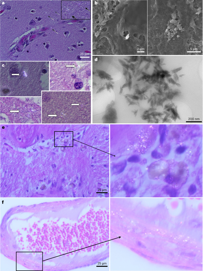
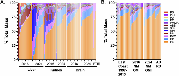
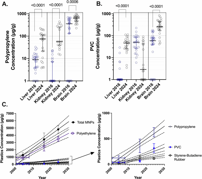
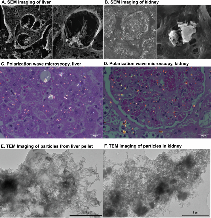
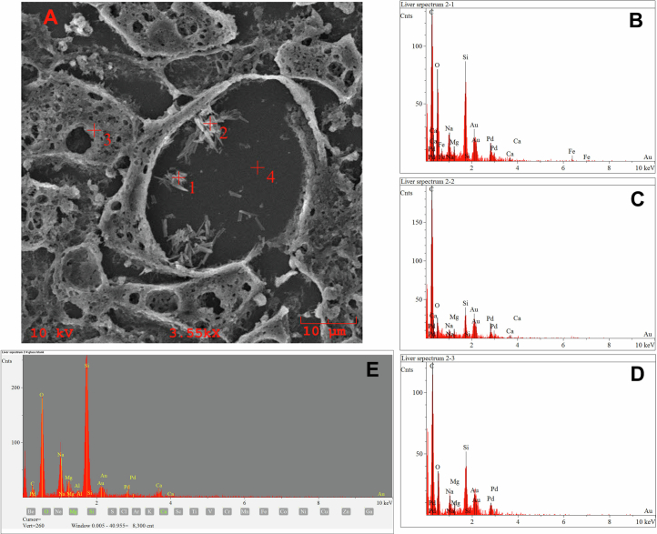
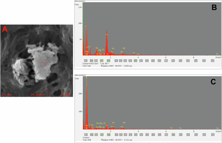
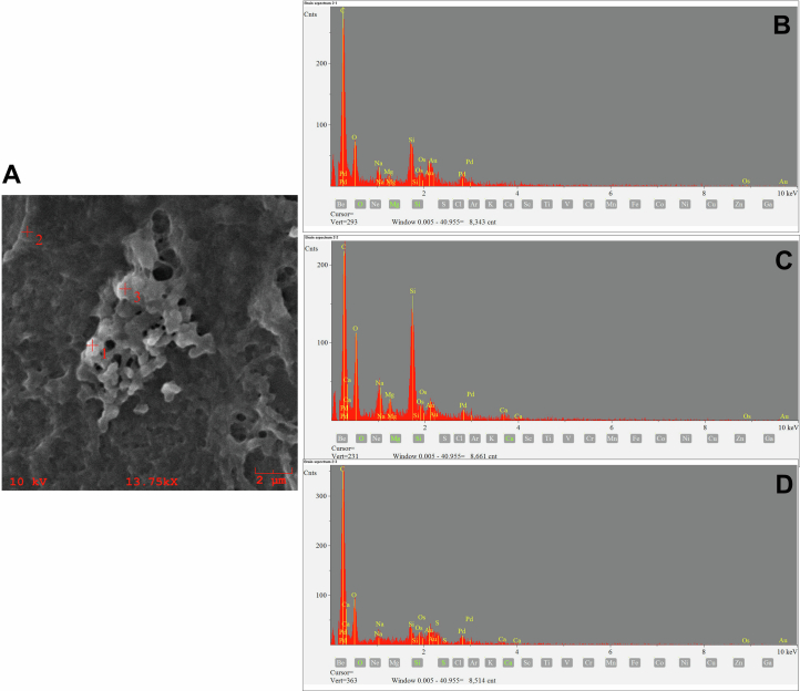
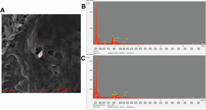
Rising global concentrations of environmental microplastics and nanoplastics (MNPs) drive concerns for human exposure and health outcomes. Complementary methods for the robust detection of tissue MNPs, including pyrolysis gas chromatography-mass spectrometry, attenuated total reflectance-Fourier transform infrared spectroscopy and electron microscopy with energy-dispersive spectroscopy, confirm the presence of MNPs in human kidney, liver and brain. MNPs in these organs primarily consist of polyethylene, with lesser but significant concentrations of other polymers. Brain tissues harbor higher proportions of polyethylene compared to the composition of the plastics in liver or kidney, and electron microscopy verified the nature of the isolated brain MNPs, which present largely as nanoscale shard-like fragments. Plastic concentrations in these decedent tissues were not influenced by age, sex, race/ethnicity or cause of death; the time of death (2016 versus 2024) was a significant factor, with increasing MNP concentrations over time in both liver and brain samples (P = 0.01). Finally, even greater accumulation of MNPs was observed in a cohort of decedent brains with documented dementia diagnosis, with notable deposition in cerebrovascular walls and immune cells. These results highlight a critical need to better understand the routes of exposure, uptake and clearance pathways and potential health consequences of plastics in human tissues, particularly in the brain.


Conflict of interest statement

Competing interests: The authors declare no competing interests.

Figures

Fig. 1
Fig. 1. Overview of total MNP concentrations from all decedent samples from liver, kidney and brain.
a, Microplastic concentrations in liver, kidney and brain decedent human samples (n = 20–28 separate participants for each timepoint; Supplementary Table 1) from the UNM OMI. Data are shown on a log10 scale, with the bar representing the group median value and 95% confidence interval. Orange-colored symbols in the 2016 brain samples were analyzed independently at Oklahoma State University. P values from Mann–Whitney tests (two-sided) indicate significant differences in samples from the same organ between 2016 and 2024 (with more comprehensive statistical treatments in Supplementary Methods—Statistical analysis). Brain MNP concentrations were significantly higher than liver and kidney, analyzed by two-way ANOVA (P < 0.0001). b, Overall distribution of 12 different polymers suggests a greater accumulation of PE in the brain relative to liver or kidney (average shown per group; see Extended Data Fig. 1 for individual data). c, PE (which was in the highest abundance and consistently had the highest confidence spectra) concentrations in all organs followed similar trends compared to total plastics (also represented as group median value and 95% confidence interval; two-sided Mann–Whitney test). d, Additional brain samples from specimens collected from 1997 to 2013 were obtained from the Duke Kathleen Price Bryan Brain Bank in North Carolina (n = 13, blue diamonds; NC), the Harvard Brain Tissue Resource Center in Massachusetts (n = 9, green diamonds; MA) and the National Institute of Child Health and Human Development Brain and Tissue Bank at the University of Maryland (n = 5, orange diamonds; MD) show lower concentrations of microplastics. Brain samples from decedents with diagnosed dementia (n = 12, purple circles) from UNM exhibit far greater MNP concentrations than brain tissues from participants without dementia from New Mexico (red thin-outline diamonds; NM). Overall linear regression trend was significantly nonzero (P < 0.0001) with an R2 = 0.3982; summary points for 2016 and 2024 normal UNM OMI brains reflect mean ± s.d. N66, nylon 66; ABS, acrylonitrile butadiene styrene; PET, polyethylene terephthalate; N6, nylon-6; PMMA, poly(methyl methacrylate); PU, polyurethane; PC, polycarbonate; PS, polystyrene.
Fig. 2
Fig. 2. Visualization of putative plastics in the brain.
a,b, Polarization wave microscopy (a, black arrows indicate refractory inclusions; inset is a digital magnification for clarity) and SEM (b, visual fields are 15.4 and 20.1 µm wide) were used to scan sections of brain from decedent human samples. c, Large (>1 µm) inclusions were not observed; additional polarization wave examples are highlighted (white arrows highlight submicron refractory inclusions). Resolution limitations of these technologies drove the use of TEM to examine the extracts from the pellets used for Py-GC/MS. d, Example TEM images resolved innumerable shard- or flake-like solid particulates following dispersion, with dimensions largely <200 nm in length and <40 nm in width. e,f, Polarization wave microscopy reveals substantially more refractile inclusions in dementia cases, especially in regions with associated immune cell accumulation (e) and along the vascular walls (f). All images were collected on a small subset of participants (n = 10 for normal brains; n = 3 for dementia cases) to provide visual evidence to support analytical chemistry.
Extended Data Fig. 1
Extended Data Fig. 1. Overall compositional outcomes, in relative proportion of the total polymer mass.
Overall compositional outcomes, in relative proportion of the total mass, are shown for (a) liver, kidney and brain samples and (b) a cross-comparison of data for brains from all cohorts (polyethylene (PE), polypropylene (PP), polystyrene (PS), acrylonitrile butadiene styrene resin (ABS), sytrene-butadiene rubber (SBR), polymethyl methacrylate (PMMA), polycarbonate (PC), polyvinyl chloride (PVC), polyurethane (PU), polyethylene terephthalate (PET), nylon-6 (N6) and nylon-6,6 (N66)). Additionally, relative proportions of polymers from Fourier transform infrared (FTIR) spectroscopic analysis in brains are shown for comparison (last 5 columns in a). In general, 4 polymers, PE, PP and PVC comprise approximately 90% of the mass of samples, with nylon being an additional major component in some samples. Each column represents a unique subject (see Supplementary Table 1 for demographic data).
Extended Data Fig. 2
Extended Data Fig. 2. Comparison of polypropylene and polyvinyl chloride in all organs across time.
Comparison of (a) polypropylene and (b) polyvinyl chloride across time and organs for NM OMI samples (see subject demographics in Supplementary Table 1). P-values shown indicate significant differences between 2016 and 2024 samples by a two-sided Mann–Whitney test. c, Simple linear regression (shown with 95% CI represented by dashed lines) was performed for total plastics, polyethylene, polypropylene, polyvinyl chloride and styrene-butadiene rubber measured in normal decedent brains from 2004 (average of east coast samples), 2016 and 2024 (NM OMI samples). Mean ± 95% CI are shown for each cluster of samples. Regression analysis for all plastics rendered a p-value < 0.0001 for each polymer, with R2 values ranging from 0.25 to 0.48.
Extended Data Fig. 3
Extended Data Fig. 3. TEM, polarization wave microscopy, and SEM images of putative microplastics from liver and kidney.
Example SEM (a,b) and polarization wave microscopy (c,d) images of decedent histological specimens and TEM images (e,f) of nanoparticulates derived from liver (left) and kidney (right). While these methods do not permit spectroscopic identification of particulate molecular composition, the bulk of particulates that were predominantly polymer as assessed by ATR–FTIR appear to be of these sizes and shapes. Energy-dispersive spectroscopy confirmed that particles were carbon-based and not mineral (Extended Data Figs. 4 and 5). Visual fields for SEM were 39.5 µm and 15.4 µm (a) and 135 µm and 9 µm (b). Example TEM images from the dispersed pellet that was derived from KOH digestion and ultracentrifugation resolved innumerable shard-like solid particulates, with dimensions largely <200 nm in length and <40 nm in width. Polarization wave microscopic images were collected on a small subset of subjects (N = 12) to provide visual evidence to support analytical chemistry.
Extended Data Fig. 4
Extended Data Fig. 4. SEM–EDS imaging of solid inclusions from the hepatic lipid droplets.
Locations of EDS are described in the SEM image (a). Particles in the droplet (1,2) render carbon-rich spectra (b,c) compared to a region (3) of hepatic tissue (d). The droplet is transected by the sectioning, and thus the background (4) reveals a silica-rich spectra consistent with the glass histology slide (e). Importantly, these particulates do not appear to be metallic or mineral. Imaging was conducted on sections from two subjects with consistent findings.
Extended Data Fig. 5
Extended Data Fig. 5. SEM–EDS imaging of solid inclusions in the kidney.
Locations of EDS are described in the SEM image (a). Renal tissue (b) displays spectra with lower relative carbon concentration than the observed particle (c). Importantly, these particulates do not appear to be metallic or mineral. Silicon and gold signals are derived from the mounting media. Imaging was conducted on sections from two subjects with consistent findings.
Extended Data Fig. 6
Extended Data Fig. 6. SEM–EDS imaging of a particulate cluster in a brain specimen.
Locations of EDS are described in the SEM image (a). The particulate regions (locations 1 and 3) exhibit a greater carbon signal in the spectra (b,d) compared to the background brain tissue (location 2; c). Importantly, these particulates do not appear to be metallic or mineral. Imaging was conducted on sections from two subjects with consistent findings.
Extended Data Fig. 7
Extended Data Fig. 7. SEM–EDS imaging of particulate in a brain specimen.
Locations of EDS are described in the SEM image (a). The background brain tissue (location 1) exhibits a lower carbon signal in the spectrum (b) compared to the particulate region (location 2; c). Importantly, these particulates do not appear to be metallic or mineral. Imaging was conducted on sections from two subjects with consistent findings.

Update of

References

    1. Thompson, R. C. et al. Lost at sea: where is all the plastic? Science304, 838 (2004). - PubMed
    1. Stubbins, A., Law, K. L., Munoz, S. E., Bianchi, T. S. & Zhu, L. Plastics in the Earth system. Science373, 51–55 (2021). - PubMed
    1. Liu, S. et al. Microplastics in three types of human arteries detected by pyrolysis-gas chromatography/mass spectrometry (Py-GC/MS). J. Hazard. Mater.469, 133855 (2024). - PubMed
    1. Marfella, R. et al. Microplastics and nanoplastics in atheromas and cardiovascular events. N. Engl. J. Med.390, 900–910 (2024). - PMC - PubMed
    1. Dong, C. D. et al. Polystyrene microplastic particles: in vitro pulmonary toxicity assessment. J. Hazard. Mater.385, 121575 (2020). - PubMed

LinkOut - more resources