. 2025 Feb 4;23(1):32.
doi: 10.1186/s12915-025-02126-w.

O-GlcNAcylation attenuates ischemia-reperfusion-induced pulmonary epithelial cell ferroptosis via the Nrf2/G6PDH pathway

Affiliations

O-GlcNAcylation attenuates ischemia-reperfusion-induced pulmonary epithelial cell ferroptosis via the Nrf2/G6PDH pathway

Liuqing Yang et al. BMC Biol. .

Abstract

Background: Lung ischemia-reperfusion (I/R) injury is a common clinical pathology associated with high mortality. The pathophysiology of lung I/R injury involves ferroptosis and elevated protein O-GlcNAcylation levels, while the effect of O-GlcNAcylation on lung I/R injury remains unclear. This research aimed to explore the effect of O-GlcNAcylation on reducing ferroptosis in pulmonary epithelial cells caused by I/R.

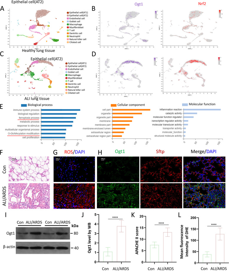
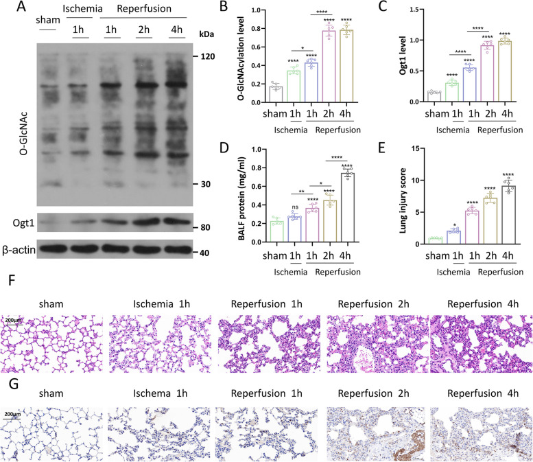
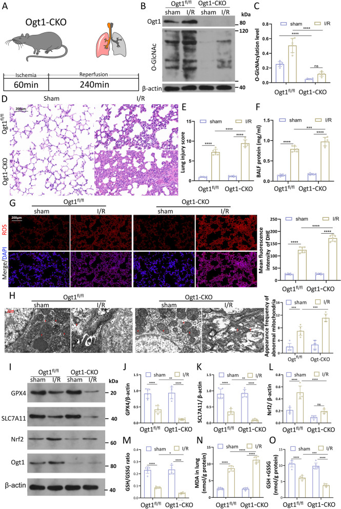
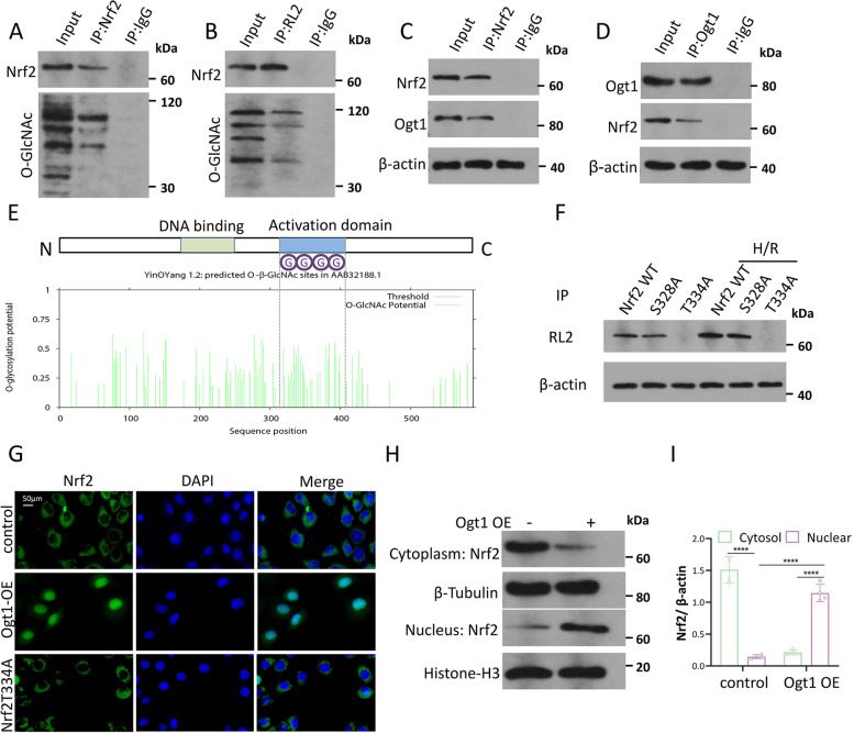
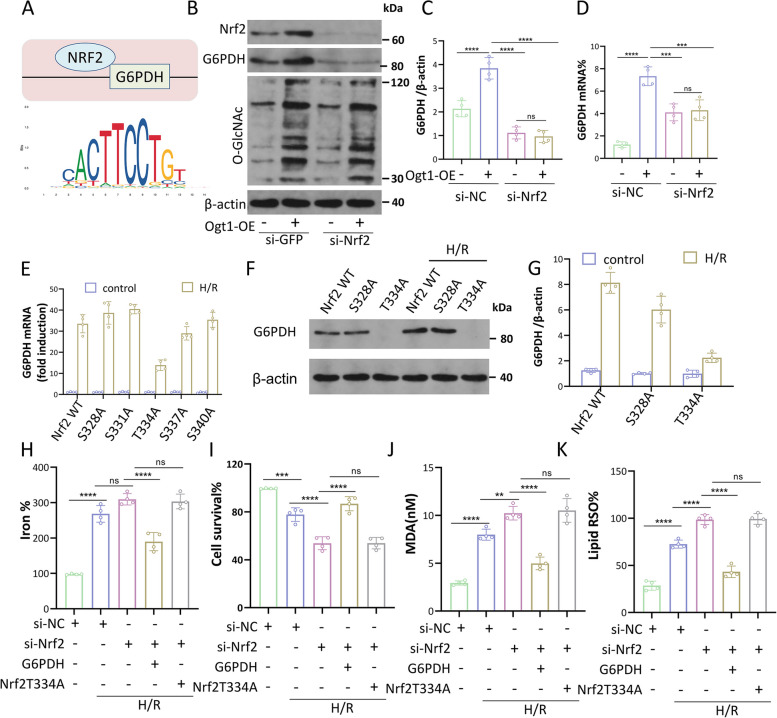
Results: First, we identified O-GlcNAc transferase 1 (Ogt1) as a differentially expressed gene in lung epithelial cells of acute lung injury/acute respiratory distress syndrome (ALI/ARDS) patients, using single-cell sequencing, and Gene Ontology analysis (GO analysis) revealed the enrichment of the ferroptosis process. We found a time-dependent dynamic alteration in lung O-GlcNAcylation during I/R injury. Proteomics analysis identified the differentially expressed proteins enriched in ferroptosis and multiple redox-related pathways based on KEGG annotation. Thus, we generated Ogt1-conditional knockout mice and found that Ogt1 deficiency aggravated ferroptosis, as evidenced by lipid reactive oxygen species (lipid ROS), malondialdehyde (MDA), Fe2+, as well as alterations in critical protein expression glutathione peroxidase 4 (GPX4) and solute carrier family 7 member 11 (SLC7A11). Consistently, we found that elevated O-GlcNAcylation inhibited ferroptosis sensitivity in hypoxia/reoxygenation (H/R) injury-induced TC-1 cells via O-GlcNAcylated NF-E2-related factor-2 (Nrf2). Furthermore, both the chromatin immunoprecipitation (ChIP) assay and the dual-luciferase reporter assay indicated that Nrf2 could bind with translation start site (TSS) of glucose-6-phosphate dehydrogenase (G6PDH) and promote its transcriptional activity. As an important rate-limiting enzyme in the pentose phosphate pathway (PPP), elevated G6PDH provided a mass of nicotinamide adenine dinucleotide phosphate (NADPH) to improve the redox state of glutathione (GSH) and eventually led to ferroptosis resistance. Rescue experiments proved that Nrf2 knockdown or Nrf2-T334A (O-GlcNAcylation site) mutation abolished the protective effect of ferroptosis resistance.

Conclusions: In summary, we revealed that O-GlcNAcylation could protect against I/R lung injury by reducing ferroptosis sensitivity via the Nrf2/G6PDH pathway. Our work will provide a new basis for clinical therapeutic strategies for pulmonary ischemia-reperfusion-induced acute lung injury.

Keywords: Ferroptosis; G6PDH; Lung ischemia–reperfusion; Nrf2; O-GlcNAcylation; Ogt1.


Conflict of interest statement

Declarations. Ethics approval and consent to participate: The procedures were conducted in accordance with the principles of Animal Care of Wuhan University (Wuhan, China) and approved by the Laboratory Animal Committee (ZN2022284). Ethical principles for clinical research of Zhongnan Hospital of Wuhan University were followed, with approval from the Medical Ethics Committee (2023138 K). All participants signed informed consent forms, and strict measures were taken to protect their privacy and data security. Consent for publication: Not applicable. Competing interests: The authors declare no competing interests.

Figures

Fig. 1
Fig. 1
The expression of Ogt1 in the lung tissues of the ALI/ARDS patient. A Cell cluster analysis showed the distribution of cell subsets in human lung tissue in the control group. Six patients with ALI/ARDS constitute the experimental group. B Uniform manifold approximation and projection (UMAP) indicated the enrichment of Ogt1 and Nrf2 genes in cell clusters in the human lung tissues of the control group. C Cell cluster analysis showed the distribution of cell subsets in human lung tissue in the ALI/ARDS group. Ten patients undergoing pulmonary benign nodule resection serve as the control group. D UMAP indicated the enrichment of Ogt1 and Nrf2 genes in cell clusters in the human lung tissues of the ALI/ARDS group. E GO enrichment analysis was conducted on the differentially expressed genes between the two groups. The analysis revealed the enrichment of signaling pathways, biological processes, and molecular functions in pulmonary epithelial cells. The X axis legend means gene number. F The representative image of HE staining lung tissues in 2 groups. 400x. G and L The representative image of immunofluorescence staining results of DHE and the corresponding quantitative analysis in 2 groups. H Immunofluorescence staining of Ogt1 expression in alveolar epithelial type II cell (AT2). I-K Comparison of Ogt1 expression and APACHE II score in lung tissues between the two groups. Data are shown as the mean ± SD. *P < 0.05, ****P < 0.0001for indicated comparisons. (Six patients with ALI/ARDS constitute the experimental group, and ten patients undergoing pulmonary benign nodule resection serve as the control group)
Fig. 2
Fig. 2
Dynamic alterations in O-GlcNAcylation after pulmonary ischemia–reperfusion injury. A-C Mice lung homogenates treated with ischemia for 1 hour, followed by reperfusion for the indicated times (1, 2 and 4 h) were used to measure the level of Ogt1 and total O-GlcNAcylated proteins. The graph displays the quantifications for each of the blots' positive bands. D The protein levels of BALF were assessed at the indicated time by the corresponding commercial kit. E Histological lung injury score was evaluated at the indicated time. F Representative images (200x) of HE staining in the lung sections at indicated time. G Chromogenic immunohistochemistry of the I/R injury lung targeting the protein Ogt1 at the indicated time. (n = 6). Data are shown as the mean ± SD. *P < 0.05, ****P < 0.0001 for indicated comparisons. BALF: Bronchoalveolar Lavage Fluid
Fig. 3
Fig. 3
Nrf2 was O-GlcNAcylated during pulmonary I/R injury in vivo, which increased the expression of its target genes. A Protein expression profiles for mice treated with or without I/R are displayed in a hierarchical clustered heatmap. B Enrichment analysis of the Kyoto Encyclopaedia of Genes and Genomes. C Nrf2–antioxidant response element pathway (Nrf2-ARE). D Mice were treated with or without I/R, and the mRNA levels of quinone oxidoreductase 1 (NQO1) (D), Glutamate Cysteine Ligase, Modifier (GCLM) (E), heme oxygenase-1 (HO-1) (F), Glutathione S-transferase (GST) (G), У-glutamylcysteine synthetase heavy(GCSH) (H) and Glucose 6 Phosphate Dehydrogenase(G6PDH) (I) were examined. J-K) G6PDH activity(J), NADPH/NADP+ ratio(K), the contents of glutathione (GSH) (M), and GSH/GSSG ratio (N) of lung tissue lysates are measured by the corresponding commercial kit. (n = 6). Data are shown as mean ± SD. *P < 0.05, ****P < 0.0001 for indicated comparisons. I/R: ischemia-reperfusion
Fig. 4
Fig. 4
Inhibition of O-GlcNAcylation mediated by Ogt1 deficiency exaggerated I/R-induced pulmonary ferroptosis in mice. A The experimental protocol's schematic representation. B, C Ogt1 conditional knockout in mice pulmonary epithelial cells was certified using WB. D Histological pictures of lung tissues stained with HE of Ogt1fl/fl and Ogt1-CKO mouse lungs treated with or without I/R. 200x. E The pathological lesion scores of the lungs in the four groups. F BALF protein content. G The immunofluorescence staining results of DHE in lung tissue. H TEM presented images and quantification of mitochondrial dysfunction. The representative mitochondria are indicated by the red arrow. I-L The expression levels of GPX4 (J), SLC7A11 (K), Nrf2 (L) in mice receiving the indicated treatment were analysed using WB and densitometry data. M-O The content of GSH/GSSG ratio(M), MDA (N), and GSH+GSSG in the lungs (O) was measured. (n = 6). Data are shown as the mean ± SD. *P < 0.05, ****P < 0.0001 for indicated comparisons. WB: Western Blot MDA: Malondialdehyde
Fig. 5
Fig. 5
Augmented O-GlcNAcylation by Ogt1 overexpression improved H/R-induced ferroptosis in TC-1 cells. A Fluorescence photos of TC-1 stained TC-1 cells, treated with H/R and Ogt1 OE separately or simultaneously. B, C Quantitative consequences of Mitochondrial membrane potential (MMP) and Cell viability were evaluated in cells with the indicated treatment. D, E BODIPY 581/591 was used to analyze, and flow cytometry was used to detect the formation of lipid ROS in TC-1 cells treated with the contextual stimuli previously described. F, G The immunofluorescence staining results of DHE in TC-1 cells. H, I Mito-FerroGreen-stained TC-1 cell fluorescence images and quantitative ferrous iron data in the mitochondria. J-M WB displayed the expression levels of GPX4 (K), SLC7A11 (L), and Nrf2(M) in TC-1 cells. N-P The content of GSH/GSSG (N), MDA (O), and GSH+GSSG ratio (P) of TC-1 cells. (n=4). Data are shown as the mean ± SD. *P < 0.05, ****P < 0.0001 for indicated comparisons
Fig. 6
Fig. 6
Ogt1 knockdown impaired H/R-induced mitochondrial function and ferroptosis in TC-1 cells. A-G Measurement of DHE fluorescence intensity (A), GSH+GSSG (B) and MDA content (C), Cell viability (D), the level of MMP (E), Fe2+(F) and the GSH/GSSG ratio in TC-1 cells treated with H/R and si-Ogt1 separately or simultaneously. (n=4). Data are shown as the mean ± SD. *P < 0.05, ****P < 0.0001 for indicated comparisons
Fig. 7
Fig. 7
O-GlcNAc transferase (Ogt1) promoted O-GlcNacylation of Nrf2 at the binding site T334, and then promoted its transnucleation. A, B Co-IP was used to verify the interaction between O-GlcNAc and Nrf2 using anti-Nrf2 (A) and Rl2 (B) with input as control.C, D Interaction between Ogt1 and Nrf2 using anti-Ogt1 (D) and anti-Nrf2 antibodies (C).E The YinOYang 1.2 server predicted the O-GlcNAcylated locations on Nrf2. F Site-directed mutagenesis produced a number of Nrf2 point mutations. After transfecting cells with an empty vector, WT, or mutant constructs of Nrf2, H/R was performed, and Co-IP was used to measure O-GlcNAcylated Nrf2.G Cytosolic and nuclear fractionation experiments of Nrf2. An immunofluorescence experiment was used to demonstrate Nrf2's subcellular localization in TC-1 cells with or without Ogt1-OE and Nrf2T334A treatment.H, I The subcellular localization of Nrf2 was detected using WB in TC-1 cells, and its quantitative graph is shown in (I). (n=4). Data are shown as the mean ± SD. *P < 0.05, **P < 0.01 for indicated comparisons. RL2: an antibody targeting O-linked N-acetylglucosamine
Fig. 8
Fig. 8
O-GlcNAcylated Nrf2 inhibited ferroptosis sensitivity during ischemia–reperfusion injury by targeting G6PDH. A Bioinformatics analysis indicated that G6PDH contains a binding site of Nrf2. B-D WB shows the expression of Nrf2, G6PDH, and global O-GlcNAcylation with si-Nrf2 and Ogt1 OE separately or simultaneously (B-C). Using qPCR, the transcription levels of G6PDH were assessed in TC-1 cells (D). E-G The transcription levels (E) and the protein expression of G6PDH (F-G) were determined in TC-1 cells with a series of point mutations of Nrf2. HK Ferroptosis signs were assessed in control TC-1 cells and cells with Nrf2 knockdown, G6PDH overexpression and Nrf2T334A separately or simultaneously after H/R: Fe2+(H), cell survival (I), MDA (J), and lipid ROS production (K). (n=4). Data are shown as the mean ± SD. *P < 0.05, **P < 0.01 for indicated comparisons.
Fig. 9
Fig. 9
Nrf2 regulated G6PDH expression by binding to its promoter region. A TC-1 cells were used to detect luciferase activity using a dual-luciferase system with the specified truncations of the G6PDH promoter. B The G6PDH binding site was predicted by bioinformatics study. C Gene G6PDH's anticipated binding site was removed, named Del-Nrf2. In TC-1 cells, luciferase activity was quantified. D Location of PBS on G6PDH, and its homologous sequence among humans, rhesus macaques, and orangutans. E, F qPCR was carried out in TC-1 cells after chromatin was immunoprecipitated using an anti-Nrf2 antibody or a negative control anti-IgG antibody (E). (F) displays its quantitative graph. G-I WB was used to identify Nrf2 expression (G), and the dual-luciferase method (H) was used to quantify G6PDH promoter activity in TC-1 cells that had both Nrf2-WT or Nrf2-T334A overexpression and simultaneous Nrf2-knockdown. Del-Nrf2 TC-1 cells were used to test the activity as well (I). (n=4). Data are shown as the mean ± SD. *P < 0.05, **P < 0.01 for indicated comparisons
Fig. 10
Fig. 10
The mechanism illustration of the involvement of Nrf2 in inhibiting ferroptosis sensitivity during I/R-induced pulmonary injury. I/R induces increased intracellular Ogt1 expression, which interacts with Nrf2 and promotes its O-GlcNacylation and transnucleation. O-GlcNAcylated Nrf2 combines with the G6PDH promoter region, increasing G6PDH expression at the transcript and protein levels. As an important rate-limiting enzyme in PPP, elevated G6PDH provides a mass of NADPH to improve the redox state of GSH, which can assist GPX4 in scavenging the mass of peroxide induced by I/R. Decreased levels of peroxide reduce cell membrane damage and resist the damage of free radicals to organelles by inhibiting the detrimental Fenton reaction between peroxide and cellular Fe2+ absorbed into cells via TFRC. Collectively, we conclude that O-GlcNAcylation attenuates I/R–induced ferroptosis in pulmonary epithelial cells via the Nrf2/G6PDH axis

References

    1. Eltzschig HK, Eckle T. Ischemia and reperfusion–from mechanism to translation. Nat Med. 2011;17(11):1391–401. - PMC - PubMed
    1. Yabuki H, Watanabe T, Oishi H, Katahira M, Kanehira M, Okada Y: Muse Cells and Ischemia-Reperfusion Lung Injury. In: Muse Cells: Endogenous Reparative Pluripotent Stem Cells. Edited by Dezawa M, vol. 1103. Cham: Springer International Publishing Ag; 2018: 293–303. - PubMed
    1. Li Y, Feng D, Wang Z, Zhao Y, Sun R, Tian D, Liu D, Zhang F, Ning S, Yao J, et al. Ischemia-induced ACSL4 activation contributes to ferroptosis-mediated tissue injury in intestinal ischemia/reperfusion. Cell Death Differ. 2019;26(11):2284–99. - PMC - PubMed
    1. Li C, Sun G, Chen B, Xu L, Ye Y, He J, Bao Z, Zhao P, Miao Z, Zhao L, et al. Nuclear receptor coactivator 4-mediated ferritinophagy contributes to cerebral ischemia-induced ferroptosis in ischemic stroke. Pharmacol Res. 2021;174: 105933. - PubMed
    1. Qin Q, Yu N, Gu Y, Ke W, Zhang Q, Liu X, Wang K, Chen M. Inhibiting multiple forms of cell death optimizes ganglion cells survival after retinal ischemia reperfusion injury. Cell Death Dis. 2022;13(5):507. - PMC - PubMed

LinkOut - more resources