. 2025 Feb 11;122(6):e2417058122.
doi: 10.1073/pnas.2417058122. Epub 2025 Feb 4.

Spatial propagation of temperate phages within and among biofilms

Affiliations

Spatial propagation of temperate phages within and among biofilms

James B Winans et al. Proc Natl Acad Sci U S A. .

Abstract

Bacteria form groups composed of cells and a secreted polymeric matrix that controls their spatial organization. These groups-termed biofilms-can act as refuges from environmental disturbances and from biotic threats, including phages. Despite the ubiquity of temperate phages and bacterial biofilms, live propagation of temperate phages within biofilms has not been characterized on cellular spatial scales. Here, we leverage several approaches to track temperate phages and distinguish between lytic and lysogenic host infections. We determine that lysogeny within Escherichia coli biofilms initially occurs within a predictable region of cell group packing architecture on the biofilm periphery. Because lysogens are generally found on the periphery of large cell groups, where lytic viral infections also reduce local biofilm structural integrity, lysogens are predisposed to disperse into the passing liquid and are overrepresented in downstream biofilms formed from the dispersal pool of the original biofilm-phage system. Comparing our results with those for virulent phages reveals that temperate phages have unique advantages in propagating over long spatial and time scales within and among bacterial biofilms.

Keywords: biofilm; dispersal; matrix; phage; spatial ecology.


Conflict of interest statement

Competing interests statement:The authors declare no competing interest.

Figures
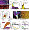
Fig. 1.
Fig. 1.
λ phages and their lysogenized hosts are mostly restricted to the outer periphery of biofilm cell groups, which is stable over days of growth; this pattern is closely dependent on host biofilm architecture. (A) WT E. coli biofilms (purple) invaded with λ phages (turquoise) for 24 h, with some cells becoming lysogenized (yellow). (B) ΔcsgBA biofilms (red) invaded with λ phages (turquoise) for 24 h, with some cells becoming lysogenized (yellow). Images in (A) and (B) are 3-dimensional renderings (for WT: 64Lx32Wx30H μm; for ΔcsgBA: 64Lx32Wx8H μm). (C and D) The frequency distributions of phages, lysogens, and total host cell populations with respect to biofilm cell packing for (C) the WT biofilm experiments and (D) the ΔcsgBA biofilm experiments. (E and F) Quantitative comparisons of the mean cell packing and the SD in cell packing near lysogenized cells in comparison with the total host bacterial population over many runs of the experiments shown in (AD). (G and H) Representative two-dimensional optical section images of experiments in which biofilms were treated as for (A and B) but were tracked for 120 h instead of 24 h. (I) Lysogen frequency over 120 h for the WT background and the ΔcsgBA background. The shaded regions around the boxplots indicate the full range of data from all replicates. (J) Phage titer over 300 min when inoculated with no bacteria, naïve E. coli, lysogenized E. coli (phage resistant and phage-adsorbing), and ΔlamB E. coli (phage-resistant, but not phage-adsorbing. The dashed line indicates the limit of detection. (K) Bacterial CFU count for naïve host E. coli and lysogenized E. coli, as well as λ phage titer over 300 min when phages are introduced to a population of only naïve hosts in shaken liquid culture. For comparison, a separate control culture of phage-naïve E. coli with no phage added is shown as well. The dashed line indicates the limit of detection.
Fig. 2.
Fig. 2.
Lysogens are overrepresented in the liquid exiting chambers because they arise in peripheral biofilm regions with low packing density. (A) Representative two-dimensional optical section of a phage-naïve WT E. coli (purple) biofilms following exposure to phages (cyan). Lysogens (yellow) have been formed around the periphery of the biofilm cell group. This image is the bottom layer of a 3-D biofilm cluster—for full data analysis that follows, the entire 3-D biofilm’s volume is imaged and quantified. (B) A heatmap showing a two-dimensional projection of cell packing measurements for the full 3-dimensional z-stack of images capturing the group of cells shown in (A). Note the reduction in cell packing as one moves from the center to the edge of the colony. (C) A comparison of the frequency of lysogens in liquid effluent exiting biofilm chambers, relative to the frequency of lysogens in regions of different cell packing. Effluent lysogen frequency is closest to that in the regions of with average cell packing <0.2 on the periphery of biofilm clusters; this peripheral, loosely packed region of biofilm clusters makes the largest contribution to the dispersal pool. (D) Measurements of distance to outer cell cluster surface for the total prey cell population, lysogens, and phages. Phages and lysogens are restricted to the outermost layers of cells with low packing density. (E) Frequency distributions of distance to outer cell group surface for the total prey population, lysogens, and phages across all experimental replicates. (F) An illustration of the dispersal assay in which biofilms are first grown in the left hand (“original”) chamber, after which the effluent from this chamber is used to inoculate a second (“downstream”) chamber to simulate dispersal from one location to another. (G) The frequency of lysogens in the original upstream chamber and the new downstream chamber for dispersal experiments with the WT E. coli strain background and the isogenic ΔcsgBA mutant that cannot produce curli matrix.
Fig. 3.
Fig. 3.
Phages released from lysogens within WT biofilms are not able to spread through the rest of the system; phages released from lysogens in a ΔcsgBA background do spread through the rest of the population due to lack of high cell packing and diffusion impedance in ΔcsgBA biofilms. Biofilms of either (A) the WT background or (D) the ΔcsgBA background were inoculated with a 1:10 mixture of λ lysogens and naïve host E. coli and grown for 72 h. Lysogens were then induced to switch to the lytic cycle by incubating the biofilm chambers for at 42° C for 40 min. (A) The population dynamics tracked over 120 h for uninfected E. coli hosts, parental lysogens, and newly derived lysogens produced by phages released during the induction treatment at 48 h. (B) Total frequency of (parental + newly derived) lysogens, and the (C) frequency of only the newly formed lysogens at 120 h in WT biofilms with, or without, lytic induction of prophages. (DF) Identical experiments as for (AC), but in this instance using the ΔcsgBA curli-null strain background for both the lysogen and the nonlysogen; without curli, phage diffusion is unimpeded, and a consequently large number of new lysogens are made that convert ~75% of the population into lysogens. (G) Quantification of phages exiting chambers of WT or ΔcsgBA biofilms following heat induction to prompt the lytic switch among prophages. Phage diffusion is reduced or eliminated in WT biofilms, hence far fewer phages exit the chambers after induction; for the ΔcsgBA background, whose biofilms do not block phage diffusion, a larger number of phages can be found exiting the chamber following lytic induction of prophages.
Fig. 4.
Fig. 4.
Phage T7 excels at rapidly exploiting available hosts in a large burst of lytic infection events. Phage λ however excels at increasing in relative abundance and self-sustaining within biofilm with period of dispersal and recolonization. Here, biofilms of WT E. coli were grown for 24 h before introduction of phage λ, T7, or a mutant of phage λ lacking Repressor; the latter phage was a control, as it is identical to λ but no longer lysogenizes hosts. After tracking local dynamics for 7 h, a dispersal cycle was implemented in which cells and phages exiting with the upstream chamber effluent were used to colonize new, sterile chambers (downstream chambers) (A) Biovolume of infected cells in original (upstream) microfluidic devices for 7 h of phage exposure to either T7, λ, or λΔcI phages (n = 4 to 8). (B) Biovolume of infected cells in new (downstream) microfluidic devices over a 96 h time course (n = 4 to 8). (C and D) Representative two-dimensional optical sections of t = 2 h of exposure to λ or T7 phages, respectively. (E) Infected cell frequency as a proportion of the whole population for T7 or λ in original (upstream) microfluidic device (n = 4). (F) Infected cell frequency in new (downstream) microfluidic devices (n = 4 to 8). (G and H) Representative two-dimensional optical sections of t = 96 h in the new microfluidic devices.

Update of

References

    1. Krause J., et al. , Living in Groups (OUP Oxford, 2002).
    1. Marras S., et al. , Fish swimming in schools save energy regardless of their spatial position. Behav. Ecol. Sociobiol. 69, 219–226 (2015). - PMC - PubMed
    1. Southern L. M., Deschner T., Pika S., Lethal coalitionary attacks of chimpanzees (Pan troglodytes troglodytes) on gorillas (Gorilla gorilla gorilla) in the wild. Sci. Rep. 11, 14673 (2021). - PMC - PubMed
    1. Couzin I. D., Krause J., James R., Ruxton G. D., Franks N. R., Collective memory and spatial sorting in animal groups. J. Theor. Biol. 218, 1–11 (2002). - PubMed
    1. Roussi A., Why gigantic locust swarms are challenging governments and researchers. Nature 579, 330 (2020). - PubMed

MeSH terms

LinkOut - more resources