Clinical Trial
. 2025 Feb 4;10(6):e186890.
doi: 10.1172/jci.insight.186890.

Interim report on engineered NK cell trial in lung cancer refractory to immune checkpoint inhibitors

Affiliations
Clinical Trial

Interim report on engineered NK cell trial in lung cancer refractory to immune checkpoint inhibitors

Miguel A Villalona-Calero et al. JCI Insight. .

Abstract

Background: Non-small cell lung cancer (NSCLC) remains the leading cause of cancer-related mortality, necessitating the exploration of alternate therapeutic approaches. Tumor-reactive or activated-by-cytokine killers (TRACK) are PD-L1+, highly cytolytic NK cells derived from umbilical cord blood NK cells and engineered to express soluble IL-15 (sIL15), and these cells show promise in preclinical studies against NSCLC.

Methods: We assessed safety, persistence, homing, and cytotoxic activity in 6 patients with advanced, refractory, and progressing NSCLC who received a low dose of unmatched, allogeneic, off-the-shelf sIL15_TRACK NK cells. We evaluated NK cell presence and persistence with droplet digital PCR (ddPCR), flow cytometry, and immunofluorescence staining.

Results: sIL15_TRACK NK cells had peak measurements at 1 hour and became undetectable 4 hours after each infusion. Cognate ligands to activating NK cell receptors were found in NSCLC. sIL15_TRACK NK cells were observed in a lung tumor biopsy 7 days after the final infusion, confirming their sustainment and tumor-homing ability. They retained cytolytic function following isolation from the lung tumor. Three of 6 patients achieved disease stabilization on repeat imaging, while the others progressed.

Conclusion: Unmatched, allogeneic, cryopreserved, off-the-shelf sIL15_TRACK NK cells express activating receptors, home to tumor sites that express their cognate ligands, and retain cytolytic activity after infusion, underscoring their potential as a therapeutic approach in solid tumors. At low doses, the therapy was safely administered and showed preliminary evidence of activity in 3 of 6 patients with advanced and progressive NSCLC. Additional dose escalation cohorts and coadministration with atezolizumab are planned.

Clinicaltrials: gov NCT05334329.

Funding: Funding was provided by CytoImmune Therapeutics and grants from the National Cancer Institute (CA266457, CA033572, and CA210087).

Keywords: Clinical trials; Innate immunity; Lung cancer; NK cells; Oncology.


Conflict of interest statement

Conflict of interest: JY and MAC are cofounders and shareholders of CytoImmune Therapeutics. MAC serves as a consultant to CytoImmune Therapeutics, a financial sponsor of this study. JY and MAC have a patent filed through City of Hope for sIL15_TRACK NK cells (US20220249564A1).

Figures

Figure 1
Figure 1. Protocol schema for first-in-human phase I trial of sIL15_TRACK NK cells in NSCLC (NCT05334329).
LD, lymphodepletion; PI, principal investigator.
Figure 2
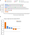
Figure 2. Depiction of patient outcomes.
(A) Swimmer plot for all 6 patients. Three patients (patients 001, 003, and 005) exhibited stable disease prior to progression. One patient (patient 001) died unexpectedly of a cardiac event with a diagnosis of COVID while on the second treatment cycle without progression. Three patients (patients 002, 004, and 006) exhibited no stabilization of disease. The swimmer plot also shows systemic palliative treatment following treatment with sIL15_TRACK NK cells. Three patients remained alive after 70, 80, and 90 weeks. (B) Waterfall plot for all 6 patients showing change in target lesions based on Response Evaluation Criteria in Solid Tumors (RECIST). The tumor volume of the target lesions in patient 001 following 1 cycle of therapy with sIL15_TRACK NK cells was reduced by approximately 12% (Supplemental Figure 2).
Figure 3
Figure 3. ddPCR assessment of sIL15_TRACK NK cells in vivo.
(A) ddPCR assessment of sIL15_TRACK NK cells present in the sera of 3 patients, as measured at 0 hour (before infusion), 1 hour (shown), and 4 hours (data not shown) following the infusion of sIL15_TRACK NK cells on days 0, 7, 14, and 21 during the first cycle of therapy at DL2. Despite weekly equivalent dosing, at times we noted a progressive albeit modest increase in the 1-hour signal from day 0 to day 21. (B) ddPCR assessment of sIL15_TRACK NK cells present in the serum of patient 005, as measured at 0 hour (before infusion), 1 hour (shown), and 4 hours (data not shown) following the infusion of sIL15_TRACK NK cells on days 0, 7, and 14 during the second cycle of therapy at DL2. Again, despite weekly equivalent dosing, a progressive albeit modest increase in the 1-hour signal from day 0 to day 14 is noted, suggesting somewhat of a cumulative effect of the dosing.
Figure 4
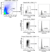
Figure 4. Flow cytometric assessment of patient lung tumor skinny needle biopsy.
The biopsy from patient 006 was collected 24 hours following the fourth (final) infusion of sIL15_TRACK NK cells and was digested to single-cell suspension using collagenase I and DNase I. Cells cocultured with K562 tumor target cells for 4 hours were also stained with antibodies against CD56 and tEGFR and assessed by flow cytometry to identify tCD56+tEGFR+ sIL15_TRACK NK cells within the lymphocyte gate. The activation and degranulation of sIL15_TRACK NK cells in the presence of the K562 tumor target cells were measured by CD107a. Frozen and thawed sIL15_TRACK NK cells cocultured without (middle) or with (bottom) K562 tumor target cells were stained as negative and positive controls for CD107a, respectively.
Figure 5
Figure 5. Immunofluorescence histochemical staining of patient lung tumor biopsy.
Utilizing the multicolor immunofluorescence histochemical staining, we examined lung tumor tissue from patient 006 on 3 occasions using DAPI (blue) for nuclear staining, pan-CK (gray) for lung parenchyma, CD45 (purple) for lymphocytes, CD57 (cyan) and CD56 (green) for NK cells, CD3 (yellow) for T cells, and EGFR (red) combined with CD56 or CD57 to identify NK cells transduced with the tEGFR-sIL15 construct. (A) FFPE tissue from a pretreatment tumor biopsy from patient 006 shows the presence of lung parenchyma (DAPI + pan-CK+) but the absence of tEGFR+ (red) transduced CD57+ or CD56+ NK cells. (B) FFPE tissue from the skinny needle tumor biopsy collected 24 hours following the fourth and final infusion of sIL15_TRACK NK cells without observable of tEGFR+ (red) transduced CD57+ (cyan) or CD56+ (green) cells. (C) FFPE tissue from an excisional tumor biopsy collected 7 days following the fourth and final infusion of sIL15_TRACK NK cells shows sIL15_TRACK NK cells identified in DAPI+CD45+ cells by coexpression of tEGFR (red) with CD57 (cyan), as marked by white arrows. (D) Selected individual markers of the boxed region shown in C are shown. Scale bars: 10 μm (AC); 10 μm (D).
Figure 6
Figure 6. NK cell–activating receptor ligands expressed on tissue from patients with NSCLC.
An excisional tumor biopsy specimen from patient 006 was assessed for the presence of the cognate ligands to NKp30 (i.e., BAG6 or B7H6), NKG2D (i.e., MICA/B), and DNAM-1 (i.e., CD112). Tissues were stained by multiplex immunofluorescence for DAPI (blue), pan-cytokeratin (CK, gray), BAG6 (cyan) B7H6 (green), MICA/B (yellow), and CD112 (red). Representative images are shown from posttreatment FFPE tumor tissue as a composite image (A), and as stained with pan-CK and DAPI (B), pan-CK and BAG6 (C), pan-CK and B7H6 (D), pan-CK and MICA/B (E), and pan-CK and CD112 (F). Scale bars: 100 μm.

References

    1. Bray F, et al. Global cancer statistics 2022: GLOBOCAN estimates of incidence and mortality worldwide for 36 cancers in 185 countries. CA Cancer J Clin. 2024;74(3):229–263. doi: 10.3322/caac.21834. - DOI - PubMed
    1. Malhotra J, et al. Risk factors for lung cancer worldwide. Eur Respir J. 2016;48(3):889–902. doi: 10.1183/13993003.00359-2016. - DOI - PubMed
    1. Molina JR, et al. Non-small cell lung cancer: epidemiology, risk factors, treatment, and survivorship. Mayo Clin Proc. 2008;83(5):584–594. doi: 10.1016/S0025-6196(11)60735-0. - DOI - PMC - PubMed
    1. Garon EB, et al. Pembrolizumab for the treatment of non-small-cell lung cancer. N Engl J Med. 2015;372(21):2018–2028. doi: 10.1056/NEJMoa1501824. - DOI - PubMed
    1. Borghaei H, et al. Nivolumab versus docetaxel in advanced nonsquamous non-small-cell lung cancer. N Engl J Med. 2015;373(17):1627–1639. doi: 10.1056/NEJMoa1507643. - DOI - PMC - PubMed

Publication types

Associated data