. 2024 Dec 31;73(6):1075-1084.
doi: 10.33549/physiolres.935368.

Eicosapentaenoic Acid Triggers Phosphatidylserine Externalization in the Erythrocyte Membrane through Calcium Signaling and Anticholinesterase Activity

Affiliations

Eicosapentaenoic Acid Triggers Phosphatidylserine Externalization in the Erythrocyte Membrane through Calcium Signaling and Anticholinesterase Activity

F H Alharthy et al. Physiol Res. .

Abstract

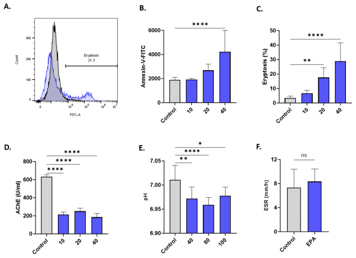
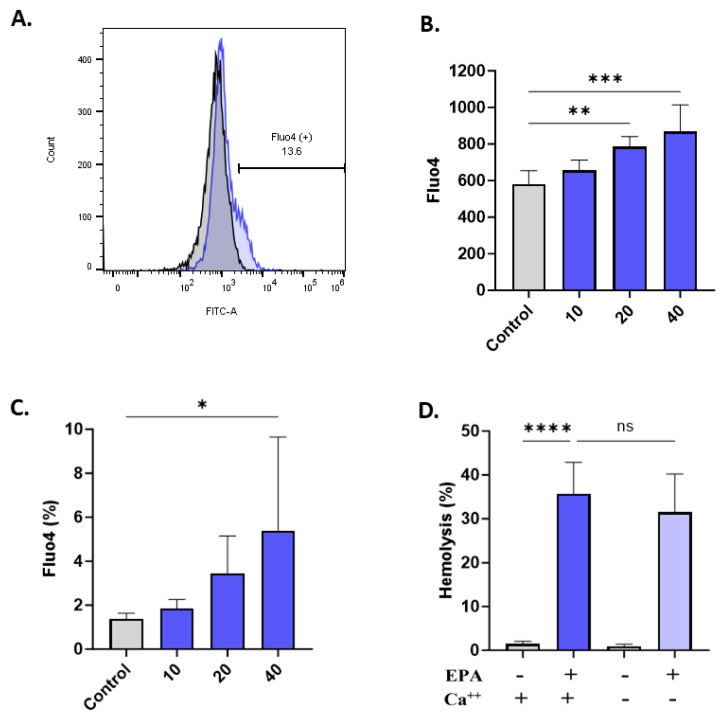
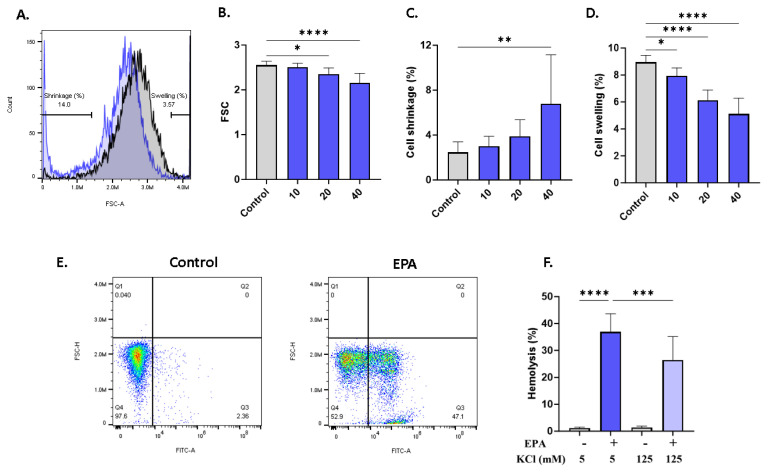
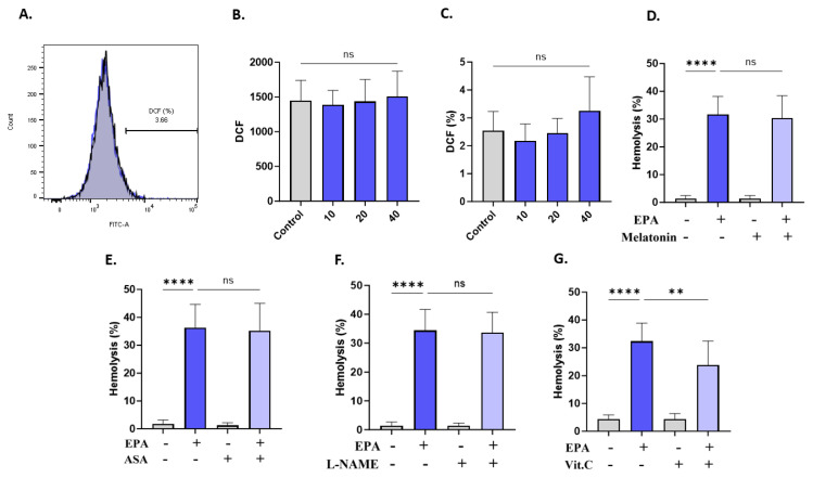
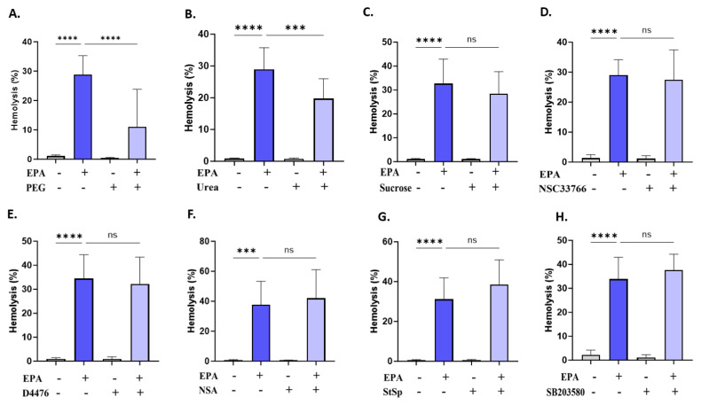
Hemolysis and eryptosis contribute to anemia encountered in patients undergoing chemotherapy. Eicosapentaenoic acid (EPA) is an omega-3 dietary fatty acid that has anticancer potential by inducing apoptosis in cancer cells, but its effect on the physiology and lifespan of red blood cells (RBCs) is understudied. Human RBCs were exposed to anticancer concentrations of EPA (10-100 ?M) for 24 h at 37 °C. Acetylcholinesterase (AChE) activity and hemolysis were measured by colorimetric assays whereas annexin-V-FITC and forward scatter (FSC) were employed to identify eryptotic cells. Oxidative stress was assessed by H2DCFDA and intracellular Ca2+ was measured by Fluo4/AM. EPA significantly increased hemolysis and K+ leakage, and LDH and AST activities in the supernatants in a concentration-dependent manner. EPA also significantly increased annexin-V-FITC-positive cells and Fluo4 fluorescence and decreased FSC and AChE activity. A significant reduction in the hemolytic activity of EPA was noted in the presence extracellular isosmotic urea, 125 mM KCl, and polyethylene glycol 8000 (PEG 8000), but not sucrose. In conclusion, EPA stimulates hemolysis and eryptosis through Ca2+ buildup and AChE inhibition. Urea, blocking KCl efflux, and PEG 8000 alleviate the hemolytic activity of EPA. The anticancer potential of EPA may be optimized using Ca2+ channel blockers and chelators to minimize its toxicity to off-target tissue. Keywords: EPA, Eryptosis, Hemolysis, Calcium, Anticancer.


Conflict of interest statement

Conflict of Interest: There is no conflict of interest.

Figures

Fig. 1
Fig. 1
EPA induces hemolysis. (A) concentration-responsive hemolytic activity of EPA (10–100 μM) in PBS and in (B) Ringer buffer. (C) K+ and (D) LDH and (E) AST activity. (F) Effect of EPA on hypotonic hemolysis. ns indicates no statistical significance, while **(p <0.01), ***(p <0.001), and ****(p <0.0001). Data were analyzed by one-way ANOVA followed by Dunnett’s correction.
Fig. 2
Fig. 2
EPA elicits eryptosis. (A) Representative histograms of annexin-V-FITC fluorescence of control (black line) and treated cells (40 μM; blue line). (B) Annexin-V-FITC fluorescence. (C) Percentage of eryptotic cells. (D) AChE activity. (E) Extracellular pH. (F) ESR. ns indicates no statistical significance, while *(p <0.05), **(p <0.01), and ****(p <0.0001). Data were analyzed by one-way ANOVA followed by Dunnett’s correction. ESR data were analyzed by Student’s t-test.
Fig. 3
Fig. 3
EPA elevates Ca2+ levels. (A) Representative histograms of Fluo4 fluorescence of control (black line) and treated cells (40 μM; blue line). (B) Fluo4 fluorescence. (C) Percentage of cells with excess Ca2+ accumulation. (D) Effect of Ca2+ availability on hemolysis. ns indicates no statistical significance, while *(p <0.05), **(p <0.01), ***(p <0.001), and ****(p <0.0001). Data were analyzed by one-way ANOVA followed by Dunnett’s correction.
Fig. 4
Fig. 4
Effect of EPA on RBC morphology. (A) Representative histograms of FSC of control (black line) and treated cells (40 μM; blue line). (B) Geomean FSC. (C) Percentage of cell shrinkage. (D) Percentage of cell swelling. (E) Dot plots of control and treated (40 μM) cells relative to FSC-H and annexin-V-FITC (annexin-V-positive cells are shown in Q3). (F) Effect of KCl (125 mM) on hemolysis. ns indicates no statistical significance, while *(p < 0.05), **(p <0.01), and ****(p <0.0001). Data were analyzed by one-way ANOVA followed by Dunnett’s correction.
Fig. 5
Fig. 5
Protective effect of vitamin C. (A) Representative histograms of DCF fluorescence of control (black line) and treated cells (40 μM; blue line). (B) DCF fluorescence. (C) Percentage of oxidized cells. Effect of (D) melatonin, (E) ASA, (F) L-NAME, and (G) vitamin C on EPA-induced hemolysis. ns indicates no statistical significance, while **(p <0.01), and ****(p <0.0001). Data were analyzed by one-way ANOVA followed by Dunnett’s correction.
Fig. 6
Fig. 6
Inhibitors of EPA-induced hemolysis. Effect of (A) PEG, (B) urea, (C) sucrose, (D) NSC23766, (E) D4476, (F) NSA, (G) StSp, and (H) SB203580 on EPA-induced hemolysis. ns indicates no statistical significance while ***(p <0.001), and ****(p <0.0001). Data were analyzed by one-way ANOVA followed by Dunnett’s correction.

References

    1. Ashfaq W, Rehman K, Siddique MI, Khan QAA. Eicosapentaenoic acid and docosahexaenoic acid from fish oil and their role in cancer research. Food Rev Int. 2020;36:795–814. doi: 10.1080/87559129.2019.1686761. - DOI
    1. Gammone MA, Riccioni G, Parrinello G, D'orazio N. Omega-3 polyunsaturated fatty acids: Benefits and endpoints in sport. Nutrients. 2019;11:1–16. doi: 10.3390/nu11010046. - DOI - PMC - PubMed
    1. Volpato M, Hull MA. Omega-3 polyunsaturated fatty acids as adjuvant therapy of colorectal cancer. Cancer Metastasis Rev. 2018;37:545–555. doi: 10.1007/s10555-018-9744-y. - DOI - PMC - PubMed
    1. Kelavkar UP, Hutzley J, Dhir R, Kim P, Allen KGD, McHugh K. Prostate tumor growth and recurrence can be modulated by the ω-6:ω-3 ratio in diet: Athymic mouse xenograft model simulating radical prostatectomy. Neoplasia. 2006;8:112–24. doi: 10.1593/neo.05637. - DOI - PMC - PubMed
    1. Sawada N, Inoue M, Iwasaki M, Sasazuki S, Shimazu T, Yamaji T, et al. Consumption of n-3 fatty acids and fish reduces risk of hepatocellular carcinoma. Gastroenterology. 2012;142:1468–75. doi: 10.1053/j.gastro.2012.02.018. - DOI - PubMed

MeSH terms