

Upright positioning facilitates the absorption of macular hole-related oedema
- PMID: 39904785
- PMCID: PMC12149008
- DOI: 10.1007/s00417-025-06757-1
Upright positioning facilitates the absorption of macular hole-related oedema
Abstract
Purpose: To investigate changes in macular hole-related oedema depending on positioning.
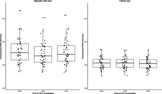
Methods: Prospective interventional study of 40 patients with primary macular hole (MH). Optical coherence tomography scanning was done at 9 a.m., 1 p.m., and 3 p.m. Between the first and second scanning, the patients were instructed to stay upright, whereas they were positioned recumbent thereafter. Automated mean retinal thickness measurements were derived from the ETDRS grid for the central, parafoveal, and perifoveal subfields. Mean ocular perfusion pressure (MOPP) was calculated for all time points. Primary endpoints were changes in MH-related oedema from 9 a.m.-1 p.m., and from 1 p.m.-3 p.m.
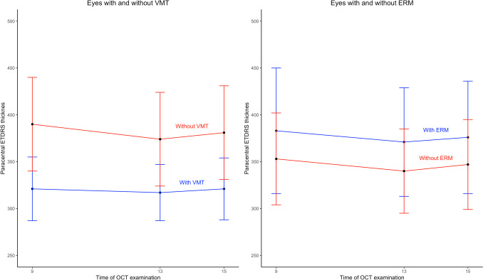
Results: In upright position from 9 a.m.-1 p.m., the mean parafoveal retinal thickness decreased from 362 μm (SD = 56) to 350 μm (SD = 51) (P < 0.001). The reduction of MH-related oedema when upright was positively correlated with a reduction in MOPP. Eyes with vitreomacular traction (VMT) exhibited significantly less reduction in MH-related oedema compared to eyes without VMT. In recumbent position from 1 p.m.-3 p.m., the mean parafoveal retinal thickness increased to 356 μm (SD = 52) (P = 0.002).
Conclusion: MH-related oedema belongs to the non-vasogenic cystoid maculopathies. The decrease in MH-related oedema when upright and its positive correlation to a reduction in MOPP is therefore unexpected. In recumbent position, the situation is reversed, and the oedema increases. This may be related to subtle leakage from the retinal capillaries. The presence of VMT seems to counteract the resolution of the oedema. In a clinical setting, upright positioning after MH surgery facilitates absorption of the oedema which is beneficial for MH closure.
Key messages: What is known: Macular hole formation is associated with cystoid macular oedema, possibly due to hydration of the outer retinal layers exposed to the hypotonic vitreous fluid. This oedema promotes the elevation of the hole edges from the retinal pigment epithelium.
What is new: Macular hole-related oedema decreases when the patients are upright and increases, in parallel with an increase in mean minimum macular hole diameter, when they are recumbent. The reduction of macular oedema is correlated with a reduction in mean ocular perfusion pressure, indicating that the oedema is influenced by subtle leakage from retinal capillaries. The results suggest that upright positioning might be beneficial in the early postoperative period of macular hole surgery.
Keywords: Circadian rhythm; Cystoid macular oedema; Diurnal rhythm; Macular hole; Macular oedema; Positioning.
© 2025. The Author(s).
Conflict of interest statement
Declarations. Ethical approval: All procedures performed in studies involving human participants were in accordance with the ethical standards of the Regional Committee for Medical Research Ethics, South-East Norway, and with the 1964 Helsinki declaration and its later amendments or comparable ethical standards. Informed consent: Written informed consent was obtained from all individual participants included in the study. Conflict of interest: All authors certify that they have no affiliations with or involvement in any organisation or entity with any financial interest (such as honoraria; educational grants; participation in speakers’ bureaus; membership, employment, consultancies, stock ownership, or other equity interest; and expert testimony or patent-licensing arrangements), or non-financial interest (such as personal or professional relationships, affiliations, knowledge or beliefs) in the subject matter or materials discussed in this manuscript. Meeting presentation: This paper was presented in part at the XXXIV Meeting of the Club Jules Gonin, Palma de Mallorca, Spain, May 2024.
Figures

References
-
- Woon WH, Greig D, Savage MD, Wilson MC, Grant CA, Mokete B, Bishop F (2015) Movement of the inner retina complex during the development of primary full-thickness macular holes: implications for hypotheses of pathogenesis. Graefe’s archive for clinical and experimental ophthalmology = Albrecht von Graefes Archiv fur klinische und experimentelle Ophthalmologie. 253:2103–2109. 10.1007/s00417-015-2951-0 - PubMed
-
- Govetto A, Sarraf D, Hubschman JP, Tadayoni R, Couturier A, Chehaibou I, Au A, Grondin C, Virgili G, Romano MR (2020) Distinctive mechanisms and patterns of Exudative Versus Tractional Intraretinal Cystoid spaces as seen with Multimodal Imaging. Am J Ophthalmol 212:43–56. 10.1016/j.ajo.2019.12.010 - DOI - PubMed
MeSH terms
LinkOut - more resources
Full Text Sources
