. 2025 Feb 4;16(1):1094.
doi: 10.1038/s41467-024-55085-1.

Regulation of senescence-associated secretory phenotypes in osteoarthritis by cytosolic UDP-GlcNAc retention and O-GlcNAcylation

Affiliations

Regulation of senescence-associated secretory phenotypes in osteoarthritis by cytosolic UDP-GlcNAc retention and O-GlcNAcylation

Donghyun Kang et al. Nat Commun. .

Abstract

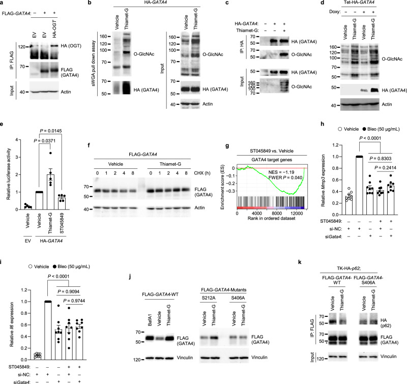
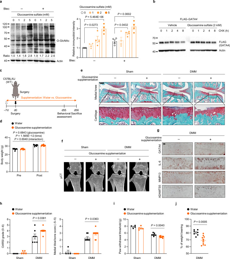
UDP-GlcNAc serves as a building block for glycosaminoglycan (GAG) chains in cartilage proteoglycans and simultaneously acts as a substrate for O-GlcNAcylation. Here, we show that transporters for UDP-GlcNAc to the endoplasmic reticulum (ER) and Golgi are significantly downregulated in osteoarthritic cartilage, leading to increased cytosolic UDP-GlcNAc and O-GlcNAcylation in chondrocytes. Mechanistically, upregulated O-GlcNAcylation governs the senescence-associated secretory phenotype (SASP) by stabilizing GATA4 via O-GlcNAcylation at S406, which compromises its degradation by p62-mediated selective autophagy. Elevated O-GlcNAcylation in the superficial layer of osteoarthritic cartilage coincides with increased GATA4 levels. The topical deletion of Gata4 in this cartilage layer ameliorates post-traumatic osteoarthritis (OA) in mice while inhibiting O-GlcNAc transferase mitigates OA by decreasing GATA4 levels. Excessive glucosamine-induced O-GlcNAcylation stabilizes GATA4 in chondrocytes and exacerbates post-traumatic OA in mice. Our findings elucidate the role of UDP-GlcNAc compartmentalization in regulating secretory pathways associated with chronic joint inflammation, providing a senostatic strategy for the treatment of OA.


Conflict of interest statement

Competing interests: The authors declare the following competing interests: Y.-J.K. and J.-H.K. are co-founders of Liflex Science. D.K., J.L., C.K., and J.-H.K. are the inventors of a patent application based on this study (South Korea patent pending, No. 10-2024-0155038). The other authors declare no competing interests.

Figures

Fig. 1
Fig. 1. The expression of UDP-GlcNAc transporters is downregulated in OA-associated chondrocytes, correlating with cytosolic UDP-GlcNAc retention.
ac Fold change (FC) heatmap of the expression levels of transporters for UDP-GlcNAc and UDP-GalNAc in human OA cartilage and OA-relevant conditions. Public transcriptome datasets analyzed were generated from (a) human OA cartilage (GSE16464, GSE43923, GSE64394, GSE113825, GSE117999, GSE178557, and GSE186220), (b) IL-1β-treated chondrocytes (GSE6119, GSE104793, and GSE163080), and (c) cartilage from OA models of mice (GSE26475, GSE53857, GSE101573, GSE110268, and GSE143447) and rats (GSE28958). d, e SA-β-Gal staining and quantification of SA-β-Gal positivity (left), representative images of immunofluorescence and quantification of BrdU incorporation (middle), and relative mRNA expression levels of CDK inhibitors or Lmnb1 (right) in (d) C28/I2 chondrocytes treated with 100 nM of doxorubicin (n = 5) or in (e) mouse chondrocytes treated with 50 μg/mL of bleomycin (n = 5). f, g Relative mRNA expression levels of transporters for UDP-GlcNAc and UDP-GalNAc in (f) C28/I2 chondrocytes treated with doxorubicin (n = 5) or in (g) mouse chondrocytes treated with bleomycin (n = 5). h Quantification of UDP-GlcNAc levels in the whole cell (left) and the ER, Golgi apparatus, and cytosol (right) of C28/I2 chondrocytes treated with vehicle, DON, or doxorubicin (n = 5). i Quantification of UDP-GlcNAc levels in the whole cell (left) and cytosol (right) of mouse chondrocytes treated with vehicle, DON, or bleomycin (n = 6). j Sulfated GAG (sGAG) release of C28/I2 chondrocytes treated with vehicle or doxorubicin measured using an sGAG assay (n = 5). k sGAG release of mouse chondrocytes treated with bleomycin (n = 5). Scale bars: d(left),e(left), 50 μm, d(middle),e(middle), 25 μm. dk Data represent means ± s.e.m. P values are from two-tailed t test (dg,j,k) or one-way ANOVA followed by Dunnett’s post-hoc test (h,i). Source data are provided as a Source Data file.
Fig. 2
Fig. 2. Stress-induced O-GlcNAcylation regulates the senescence-associated secretory phenotype of chondrocytes in osteoarthritic cartilage.
a Sections of human OA cartilage stained with Alcian blue and immunostained for O-GlcNAc and p16INK4a. Representative co-immunofluorescence images of O-GlcNAc and p16INK4a. b Pie charts showing the O-GlcNAc or p16INK4a positivity from immunofluorescence in undamaged and damaged regions of human OA cartilage (n = 6). ce Quantification of SA-β-Gal positivity and western blot for global O-GlcNAcylation in primary chondrocytes from (c) humans or (d,e) mice, treated with vehicle or (c) doxorubicin (n = 4), (d) bleomycin (n = 5), or (e) H2O2 (n = 5). f, g Immunofluorescence of γ-H2AX and SA-β-Gal staining in chondrocytes treated with bleomycin (50 μg/mL) for 24 h, followed by treatment with vehicle (DMSO) or ST045849 (20 μM) for 72 h. (f) Representative images and (g) quantification of γ-H2AX and SA-β-Gal positivity (n = 5, 3 respectively). h Relative mRNA levels of senescence markers in chondrocytes treated with bleomycin, followed by treatment with vehicle or ST045849 (n = 5). i Western blot of p16INK4a, GATA4, and SASPs in primary human chondrocytes treated with doxorubicin for 14 days. The asterisks indicate active form of the MMPs (n = 4). j Immunohistochemistry of GATA4 and SASP factors in human OA cartilage (n = 3). k Representative images (top) and pie charts (bottom) of O-GlcNAc or GATA4 positivity in human OA cartilage (n = 6). l Relative mRNA levels of SASP factors in chondrocytes treated with bleomycin for 24 h followed by treatment with vehicle or ST045849 for five days (n = 8). m GSEA of RNA-seq data using “Degradation of the extracellular matrix” gene set in chondrocytes treated with bleomycin, followed by treatment with vehicle or ST045849. Scale bars: a(top),f(bottom),j, 50 μm, a(bottom),f(top),k, 25 μm. ce,g,h,l Data represent means ± s.e.m. P values are from two-tailed t test (ce), two-way ANOVA followed by Tukey’s post-hoc test (g,h,l), or one-tailed permutation test corrected for multiple comparisons with FWER (m). Source data are provided as a Source Data file.
Fig. 3
Fig. 3. The O-GlcNAcylation of GATA4 prevents its degradation via selective autophagy, driving the expression of SASPs.
a Co-immunoprecipitation of FLAG-tagged GATA4 with HA-tagged OGT in HEK293T (n = 3). b Immunoblot of pull-down using sWGA-conjugated agarose beads with or without thiamet-G treatment in HEK293T (n = 3). c Immunoblot of O-GlcNAcylation of HA-tagged GATA4 in HEK293T treated with vehicle or thiamet-G (n = 3). HA-tagged proteins were pulled down using anti-HA antibodies. MG132 (10 µM) was applied for 6 h before lysis. d Immunoblot of GATA4 in HEK293T transfected with Tet-HA-GATA4 and treated with Doxy (1 µg/mL) or left untreated, with or without thiamet-G (10 µM) for 60 h (n = 3). e GATA reporter gene assay in HEK293T transfected with TK promoter-driven empty vector (EV) or HA-GATA4, followed by treatment with vehicle, thiamet-G, or ST045849 for 36 h (n = 5). f CHX chase analysis in HEK293T-FLAG-GATA4 treated with vehicle or thiamet-G (20 μM) (n = 3). g GSEA of RNA-seq data from chondrocytes treated with bleomycin for 24 h, followed by vehicle or ST045849 for five days, using “GATA4 target genes” gene set. h, i Relative mRNA levels of (h) Mmp3 and (i) Il6 in chondrocytes treated with bleomycin for 24 h followed by transfection with control or Gata4 siRNAs, and treatment with vehicle or ST045849 for five days (n = 8). j Western blot in HEK293T transfected with FLAG-tagged GATA4 WT or mutants (S212A or S406A) with or without thiamet-G (20 µM) for 48 h (n = 3). The Bafilomycin A1 (BafA1) treatment condition blocks autophagy-mediated protein degradation. k Co-immunoprecipitation of FLAG-tagged GATA4 WT or S406A mutant with HA-tagged p62 in HEK293T (n = 3). Cells were treated with vehicle or thiamet-G (20 µM, 48 h) and BafA1 (50 nM, 24 h). The asterisk indicates IgG heavy chains. e,h,i Data represent means ± s.e.m. P values are from two-tailed t test (e), one-tailed permutation test corrected for multiple comparisons with FWER (g), or two-way ANOVA followed by Tukey’s post-hoc test (h,i). Source data are provided as a Source Data file.
Fig. 4
Fig. 4. The topical deletion of Gata4 in the superficial zone of the cartilage abolishes SASP and OA development in mice.
a Immunohistochemistry of O-GlcNAc and GATA4 in the superficial layer of the articular cartilage from 20-week-old mouse knee joints (n = 3). b Thirteen-week-old Gata4fl/fl; Col2a1-CreERT2 mice injected with vehicle or TMX received sham or DMM surgery. Joint sections were stained with safranin O, fast green, and hematoxylin, with insets magnified in the bottom row. c Cartilage destruction, subchondral bone sclerosis, osteophyte formation, and synovial inflammation were scored (n = 8, 4 for sham; n = 15, 10 for DMM). d The percentage of weight placed on the DMM-operated limb versus the contralateral limb of Gata4fl/fl; Col2a1-CreERT2 mice injected with vehicle or TMX, determined using a static weight bearing test (n = 15, 10 respectively). e Thirteen-week-old Gata4fl/fl; Prg4-CreERT2 mice injected with vehicle or TMX received DMM surgery. Joint sections were stained with safranin O, fast green, and hematoxylin, with insets magnified below and on the right. f Cartilage destruction, subchondral bone sclerosis, osteophyte formation, and synovial inflammation were scored (n = 10, 9 respectively). g Representative μCT images of sham- or DMM-operated Gata4fl/fl; Prg4-CreERT2 mice injected with vehicle or TMX. h Immunohistochemistry of MMP3, MMP13, and IL-6 in cartilage sections from DMM-operated Gata4fl/fl; Prg4-CreERT2 mice injected with vehicle or TMX (n = 3). i The percentage of weight placed on the DMM-operated limb versus the contralateral limb of Gata4fl/fl; Prg4-CreERT2 mice injected with vehicle or TMX, determined using a static weight bearing test (n = 10, 9 respectively). j Electronic Von Frey test in the ipsilateral hindpaw of sham- or DMM-operated Gata4fl/fl; Prg4-CreERT2 mice injected with vehicle or TMX (n = 10, 9 for sham; n = 10, 9 for DMM). Scale bars: a,h, 25 μm, b, 500 μm, e, 200 μm. c,d,f,i,j Data represent means ± s.e.m. P values are from Kruskal–Wallis test followed by two-tailed Mann–Whitney U test (c), two-tailed t test (d,i,j), or Mann–Whitney U test (f). Source data are provided as a Source Data file.
Fig. 5
Fig. 5. Pharmacological modulation of O-GlcNAcylation regulates GATA4 and affects post-traumatic OA.
a Twelve-week-old WT C57BL/6 J mice underwent DMM surgery followed by weekly IA injection of vehicle or thiamet-G. Joint sections were stained with safranin O, fast green, and hematoxylin, with insets magnified below and on the right. b Cartilage destruction and synovial inflammation were scored (n = 8, 9 respectively). c The percentage of weight placed on the DMM-operated limb versus the contralateral limb of the WT mice injected with vehicle or thiamet-G, determined using a static weight bearing test (n = 8, 9 respectively). d GSEA of the “Upregulated genes in OA” gene set in chondrocytes treated with bleomycin for 24 h followed by vehicle or ST045849 treatment for five days. e Twelve-week-old WT C57BL/6 J mice received DMM surgery followed by weekly IA injection of vehicle or ST045849 using a hydrogel-based drug delivery system. Joint sections were stained with safranin O, fast green, and hematoxylin, with insets magnified on the right. fh Immunohistochemistry of (f) GATA4 and cartilage matrix neoepitopes (C-telopeptide of type II collagen, CTX-II and aggrecan neoepitope, NITEGE), (g) MMP3, MMP13, IL-6, (h) senescence markers (p16INK4a and HMGB1), and HGF in cartilage sections from DMM-operated WT mice followed by IA injection with vehicle or ST045849 (n = 3). i Cartilage destruction, subchondral bone sclerosis, osteophyte formation, and synovial inflammation were scored (n = 10). j Percentage of weight placed on the DMM-operated limb versus the contralateral limb of WT mice injected with vehicle or ST045849, determined using a static weight bearing test (n = 10). k Electronic Von Frey test in ipsilateral hindpaw of sham- or DMM-operated WT mice IA-injected with vehicle or ST045849 using a hydrogel-based drug delivery system (n = 10). Scale bars: a,e, 200 μm, fh, 25 μm. b,c,ik Data represent means ± s.e.m. P values are from two-tailed Mann–Whitney U test (b,i), two-tailed t test (c,j,k), or one-tailed permutation test corrected for multiple comparisons with FWER (d). Source data are provided as a Source Data file.
Fig. 6
Fig. 6. Excessive glucosamine supplementation enhances O-GlcNAcylation and GATA4 stability, aggravating post-traumatic OA in mice.
a Western blot of global protein O-GlcNAcylation in primary mouse chondrocytes treated with or without bleomycin for 24 h, followed by varying doses of glucosamine sulfate for 72 h (left) and quantification of O-GlcNAcylation levels (right) (n = 4). b CHX chase analysis in HEK293T-FLAG-GATA4 treated with vehicle or glucosamine sulfate (2 mM) (n = 3). c Schematic illustration of dietary glucosamine sulfate supplementation in the post-traumatic OA model of WT mice. Twelve-week-old WT C57BL/6 J mice were provided with drinking water with or without glucosamine sulfate (5 mg/mL), starting seven days prior to the sham or DMM operation. d Body weight of the DMM-operated mice pre- and post-supplementation of glucosamine sulfate (n  =  7, 10 respectively). e Joint sections were stained with safranin O, fast green, and hematoxylin, with insets magnified in the bottom row. f Representative μCT images of sham- or DMM-operated WT mice provided with or without glucosamine sulfate. g Immunohistochemistry of GATA4, IL-6, MMP13, and ADAMTS5 in cartilage sections from DMM-operated WT mice provided with or without glucosamine sulfate (n = 3). h Cartilage destruction and subchondral bone sclerosis were scored (n = 4, 5 for sham; n = 7, 10 for DMM). i Electronic Von Frey test in the ipsilateral hindpaw of sham- or DMM-operated WT mice provided with or without glucosamine sulfate (n = 4, 5 for sham; n = 7, 10 for DMM). j The percentage of weight placed on the DMM-operated limb versus the contralateral limb of the mice, determined using a static weight bearing test (n = 7, 10 respectively). Scale bars: e, 200 μm, g, 25 μm. a,d,hj Data represent means ± s.e.m. P values are from two-way ANOVA followed by Dunnett’s post-hoc test (a), two-way ANOVA followed by Bonferroni post-hoc test (d), two-tailed Mann–Whitney U test (h), or two-tailed t test (i,j). Source data are provided as a Source Data file.

References

    1. Hunter, D. J., March, L. & Chew, M. Osteoarthritis in 2020 and beyond: a Lancet Commission. Lancet396, 1711–1712 (2020). - PubMed
    1. Jeon, O. H. et al. Local clearance of senescent cells attenuates the development of post-traumatic osteoarthritis and creates a pro-regenerative environment. Nat. Med23, 775–781 (2017). - PMC - PubMed
    1. Coryell, P. R., Diekman, B. O. & Loeser, R. F. Mechanisms and therapeutic implications of cellular senescence in osteoarthritis. Nat. Rev. Rheumatol.17, 47–57 (2021). - PMC - PubMed
    1. Kang, D. et al. Stress-activated miR-204 governs senescent phenotypes of chondrocytes to promote osteoarthritis development. Sci Transl Med11, 10.1126/scitranslmed.aar6659 (2019). - PubMed
    1. Heinegard, D. & Saxne, T. The role of the cartilage matrix in osteoarthritis. Nat. Rev. Rheumatol.7, 50–56 (2011). - PubMed

MeSH terms

Substances

LinkOut - more resources