. 2025 Feb 4;16(1):68.
doi: 10.1038/s41419-025-07399-1.

CDCA7 enhances STAT3 transcriptional activity to regulate aerobic glycolysis and promote pancreatic cancer progression and gemcitabine resistance

Affiliations

CDCA7 enhances STAT3 transcriptional activity to regulate aerobic glycolysis and promote pancreatic cancer progression and gemcitabine resistance

Dijie Zheng et al. Cell Death Dis. .

Abstract

Cell division cycle associated 7 (CDCA7) plays a role in various malignancies, especially pancreatic cancer (PC). However, its expression pattern and functional significance in PC require further research. Therefore, this study aimed to investigate CDCA7 expression levels and biological functions in PC using in vitro and in vivo experiments. Western blotting, immunohistochemistry, and real-time polymerase chain reaction were performed to detect CDCA7 expression in PC cells and tissues. Additionally, the biological functions of CDCA7 were assessed using cell proliferation, wound healing, and Transwell assays. CDCA7 overexpression promoted PC cell proliferation, migration, and invasion, and increased resistance to the chemotherapy drug gemcitabine, possibly through enhanced aerobic glycolysis. Additionally, immunoprecipitation assay showed that CDCA7 interacted with STAT3 protein and affected the transcriptional regulation of hexokinase 2. Conclusively, targeting CDCA7 might be a promising therapeutic strategy to increase gemcitabine sensitivity by inhibiting glycolysis in PC cells.


Conflict of interest statement

Competing interests: The authors declare no competing interests. Ethics approval and consent to participate: All procedures followed were in accordance with the ethical standards of the Ethical Committee of the Affiliated Hospital of Guizhou Medical University. All institutional and national guidelines for the care and use of laboratory animals were followed. Consent to publication: All authors have agreed to publish this manuscript.

Figures

Fig. 1
Fig. 1. CDCA7 is highly expressed in PC tissues and cells.
A Analysis of datasets (GSE16515, GSE15471, GSE28735) from the GEO database showed that CDCA7 has a higher expression in PC tissues than in normal or non-tumor tissues. B CDCA7 mRNA expression levels were analyzed in 179 PC tissue samples and 171 normal pancreatic tissue samples using data from the GTEx and TCGA databases. C, D qRT-PCR and western blotting were performed to detected CDCA7 mRNA and protein levels, respectively, in PC tissues and para-cancerous tissues. E, F qRT-PCR and western blotting detected CDCA7 mRNA and protein expression levels, respectively, in HPNE and PC cells. G IHC confirmed CDCA7 expression levels in PC tissue and adjacent tissue. H Kaplan-Meier survival curves were generated to elucidate the survival outcomes in patients with PC with varying CDCA7 expression levels. *P < 0.05, **P < 0.01, ***P < 0.001.
Fig. 2
Fig. 2. CDCA7 promotes the proliferation, migration, and invasion of PC cells.
AC Effects of CDCA7 overexpression and knockdown on the proliferation of PC cells, detected using CCK-8 assay (A), plate clone (B), EDU (C) (scale bar: 50 μm). D, E Wound healing and Transwell assays were used to detect changes in the migratory and invasive abilities of PC cells following CDCA overexpression or knockdown. FH Establishment of xenograft tumor model, F Resection of subcutaneous tumors 42 days after transplantation of PC cells G Tumor weight. H Tumor volume. I, J IHC was performed to detect Ki-67 and PCNA expression in tumor tissues. *P < 0.05, **P < 0.01, ***P < 0.001.
Fig. 3
Fig. 3. CDCA7 enhances glycolysis in PC cells and promotes gemcitabine resistance.
A, B Samples were stratified into high (≥ 50%) and low (< 50%) expression groups based on CDCA7 expression levels, and single-gene Gene Set Enrichment Analysis (GSEA) was conducted to identify pathways associated with CDCA7 upregulation and downregulation. CE Measurement of lactate (C), glucose (D) and ATP (E) levels in PC cells following CDCA7 overexpression or knockdown. F, G Extracellular acidification rate (ECAR) indicates aerobic glycolysis level in PC cells following CDCA7 overexpression or knockdown. H CCK8 assay was performed detects the sensitivity of PC cells in the control, CDCA7 overexpression, and CDCA7 + 2-DG groups to gemcitabine. I Establishment of xenograft tumor model and isolation of the subcutaneous tumor 42 days later. J, K Tumor weight and volume. L IHC was performed to detect Ki-67 and PCNA in tumor tissues in each group. *P < 0.05, **P < 0.01, ***P < 0.001.
Fig. 4
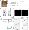
Fig. 4. Interaction between CDCA7 and STAT3.
A Detection of different protein bands using silver staining method following CDCA7 overexpression in PANC-1 cells. B Mass spectrometry analysis of five proteins coprecipitated with CDCA7. C A distinct CDCA7 polypeptide was identified from the protein lysate of anti-CDCA7 immunoprecipitated PANC-1 cells using two-dimensional LC-MS/MS mass spectrometry. D Co-immunoprecipitation assay to detect the interaction between CDCA7 and STAT3. E CDCA7 co-localizes with STAT3 in PC cells. F Design of CDCA7 and STAT3 mutants. G Immunoprecipitation and western blotting were performed to detect the interaction between flag-tagged truncated CDCA7 protein and his-tagged STAT3 protein in HEK 293 T. *P < 0.05, **P < 0.01, ***P < 0.001.
Fig. 5
Fig. 5. CDCA7 promotes PC cell progression and gemcitabine resistance by targeting STAT3 to regulate glycolysis levels.
PANC-1 and MIA PaCa-2 were transfected with or without plasmids to generate Vector, CDCA7 overexpression, and CDCA7 + sh-STAT3 groups. A CCK-8, (B) plate clone, (C) EDU assays (scale bar: 50 μm). D Wound healing (scale bar: 200 μm) and (E) Transwell assays. FH Detection of extracellular glucose and lactate levels and intracellular ATP levels in PC cells. I CCK-8 assay was performed to detect the sensitivity of each group to gemcitabine. *P < 0.05, **P < 0.01, ***P < 0.001.
Fig. 6
Fig. 6. CDCA7-STAT3 axis enhances HK2 expression and promotes glycolysis.
A Western blotting detected changes in key enzymes during glycolysis. B Correlation between CDCA7 and HK2 in the GEPIA database. C HK2 mRNA expression in PC tissue samples and normal pancreatic tissue samples using data from the GTEx and TCGA databases. D, E qRT-PCR and western blotting were performed to detect HK2 expression after CDCA7 knockdown or overexpression. F, G HK2 expression was detected in CDCA7 overexpressing cells transfected with or without sh-HK2 using qRT-PCR and western blotting. H, I Western blotting and qRT-PCR were performed detect HK2 expression following STAT3 knockdown or overexpression. J CHIP-qPCR detected STAT3 and HK2 gene. K Luciferase activity was detected PC cells co-transfected with CDCA7 and sh-STAT3. L STAT3 phosphorylation level was detected using western blotting. M Western blotting was performed to detect Whole cell lysates (WCLs) and nuclear expression of STAT3 protein. *P < 0.05, **P < 0.01, ***P < 0.001.
Fig. 7
Fig. 7. HK2 downregulation reverses STAT3-mediated PC progression.
AC CCK-8, plate cloning, and EDU assays were performed to examine cell proliferation in each group. DG Migratory and invasive abilities of cells in each group were determined using scratch and Transwell assays. HJ Lactate (H), glucose (I), and ATP levels (J) in each group. K Aerobic glycolysis was assessed in each group, based on ECAR. L CCK-8 assay was performed to examine the resistance of PC cells to gemcitabine in each group. *P < 0.05, **P < 0.01, ***P < 0.001.

References

    1. Siegel RL, Giaquinto AN, Jemal A. Cancer statistics, 2024. CA Cancer J Clin. 2024;74:12–49. - PubMed
    1. Moore A, Donahue T. Pancreatic Cancer. JAMA 2019;322:1426. - PMC - PubMed
    1. Rahib L, Smith BD, Aizenberg R, Rosenzweig AB, Fleshman JM, Matrisian LM. Projecting cancer incidence and deaths to 2030: the unexpected burden of thyroid, liver, and pancreas cancers in the United States. Cancer Res. 2014;74:2913–2921. - PubMed
    1. Conroy T, Pfeiffer P, Vilgrain V, Lamarca A, Seufferlein T, O’Reilly EM, et al. Pancreatic cancer: ESMO Clinical Practice Guideline for diagnosis, treatment and follow-up. Ann Oncol. 2023;34:987–1002. - PubMed
    1. Tang R, Xu J, Wang W, Meng Q, Shao C, Zhang Y, et al. Targeting neoadjuvant chemotherapy-induced metabolic reprogramming in pancreatic cancer promotes anti-tumor immunity and chemo-response. Cell Rep Med. 2023;4:101234. - PMC - PubMed

MeSH terms